Expand ("E")   Collapse

Age: 55

Sex: Male

Indication: Abdominal pain, recent appendectomy, history of lupus

Save ("V")

Case #1


Findings

  • Lower chest
    • Small right and trace left pleural effusions
    • Mild dependent atelectasis
  • Abdomen/Pelvis
    • Multifocal submucosal edema/hyperenhancement from the esophagus through the rectum with most severe involvement of the duodenum and proximal colon
    • Dilated, fluid-filled lower thoracic esophagus
    • Surgical changes of recent appendectomy with small volume abdominopelvic ascites, particularly in the right paracolic gutter
    • Periportal edema in the liver
    • Subcentimeter hepatic hypodensities, which are too small to characterize
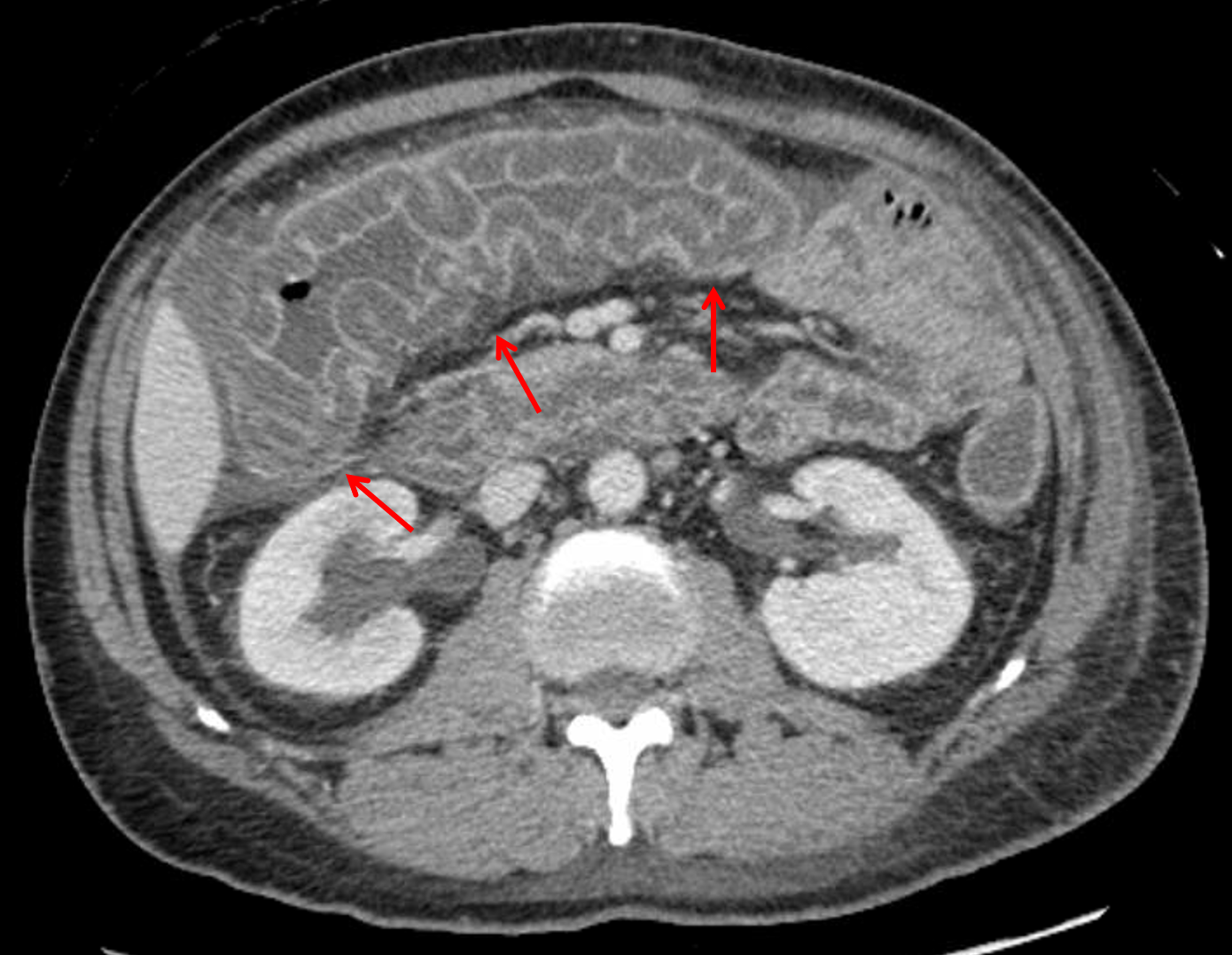
    • Mild fullness of both renal collecting systems
    • Urothelial thickening and enhancement of the bilateral ureters, particularly distally
    • Diffuse mesenteric edema
    • Trace locules of free intraperitoneal air, including adjacent to the hepatic dome and the left portal vein
  • MSK
    • No acute osseous findings
    • Slight retrolisthesis of L5 on S1
    • Body wall edema


Diagnosis

  • Small vessel vasculitis

Sample Report

Nonspecific multifocal submucosal edema throughout the bowel from the esophagus to the rectum with severe edema of the duodenum and proximal colon. In this patient with lupus, these findings are particularly concerning for a small vessel vasculitis. Infectious/inflammatory enteritis/colitis is also on the differential, but considered less likely given the distribution and discontinuous involvement.

Surgical changes of appendectomy without loculated intraabdominal collection. Trace intraperitoneal free air is likely postsurgical.

Distended, fluid-filled lower thoracic esophagus, which places the patient at risk for aspiration.

Findings suggestive of volume overload, including small pleural effusions, mesenteric edema, small volume abdominopelvic ascites, and body wall edema.

Mild fullness of the bilateral renal collecting systems with urothelial thickening and enhancement of both ureters. While this could be secondary to adjacent inflammation, recommend correlation with urinalysis if there is clinical concern for urinary tract infection.


Discussion

  • Lupus can affect any and every organ system with pathologies resulting from immune complex deposition and vasculopathy. The reference listed below gives a good overview of the spectrum of conditions associated with lupus from a radiologist’s perspective
  • When the bowel is involved, it is most often the result of small vessel disease (which is not usually directly visualized on imaging) with the SMA distribution being most commonly involved
  • Make sure to think about vasculitis and angioedema when you see segmental submucosal edema, particularly when the patient story is not typical for enteritis/colitis or inflammatory bowel disease


Images

Lupus vasculitis: marked submucosal edema at the hepatic flexure and transverse colon resulting in a thumbprinting appearance of the mucosa (red arrows).


📣 Feedback?

⌨️  Keyboard Shortcuts ("K")

Call Preparation

Prepare for call efficiently with interactive cases, sample reports, and annotated images.

1,400+ cases  91.25 CME

Call Simulator

   Neuro CT

250 cases

  Neuro MRI

146 cases

   Peds Radiographs

372 cases

   MSK Radiographs

281 cases

   Chest CT

84 cases

   Chest Radiographs

97 cases

   KUB

43 cases

   Body CT

140 cases

   Ultrasound

75 cases

Call Simulator

• Shuffle cases from our courses to simulate the mix of a call shift.
• Submit your own report before reviewing the case write-up.

Neuro Fellowship

Reviews of neuro topics with clinical pearls, differentials, and in-depth discussions.

131 cases  13.1 CME

   Brain Tumors

105 cases

   Neurodegenerative

13 cases

   Congenital Hearing Loss

13 cases

Call Preparation

   Neuro CT

250 cases

   Neuro MRI

146 cases

   Peds Radiographs

372 cases

   MSK Radiographs

281 cases

   Ultrasound

75 cases

   Chest CT

84 cases

   Chest Radiographs

97 cases

   Body CT

127 cases

   KUB

43 cases

Neuro Fellowship

   Brain Tumors

105 cases

   Neurodegenerative

13 cases

Call Simulator

• Shuffle cases from our courses.
• Submit your own report.

CME

• Earn up to 91 CME.
• Claim your CME to receive a certificate.

Call Simulator

• Shuffle cases from our courses to simulate the mix of a call shift.
• Submit your own report before reviewing the case write-up.

CME

• Earn up to 91 CME by completing cases in our radiology courses.
• Claim your CME to receive a certificate.

On Call

Quick references created specifically to help while on call.

  Peds Normals by Age

Reference database of normal imaging from birth to age 16

   Incidental Findings

Summary of consensus guidelines for managing incidental CT findings

   Media Index

Index of select illustrations & videos from our courses

  Neuro CT Mimics

Visual reference for common mimics of pathology on CT

On Call

  Peds Normals by Age

Reference database of normal imaging from birth to age 16

   Incidental Findings

Summary of consensus guidelines for managing incidental CT findings

   Media Index

Index of select illustrations & videos from our courses

  Neuro CT Mimics

Visual reference for common mimics of pathology on CT

Anatomy

Labelled radiographs and CT/MRI series teaching anatomy with a level of detail appropriate for medical students and junior residents.

   Pelvis

Pelvic MRI anatomy

   Chest

Chest radiograph & CT anatomy

   Body

Abdominal CT anatomy

   Cardiac

Cardiac CT anatomy

   Brain

Brain & calvarial anatomy on CT/MRI

   Cranial Nerves

Cranial nerves on MRI

   Shoulder

Shoulder MRI anatomy

   Knee

Knee MRI anatomy

   Temporal Bone

Resident/fellow-level anatomy

Anatomy

   Pelvis

Pelvic MRI anatomy

   Chest

Chest radiograph & CT anatomy

   Body

Abdominal CT anatomy

   Cardiac

Cardiac CT anatomy

   Brain

Brain & calvarial anatomy on CT/MRI

   Cranial Nerves

Cranial nerves on MRI

   Shoulder

Shoulder MRI anatomy

   Knee

Knee MRI anatomy

   Temporal Bone

Resident/fellow-level anatomy

Neuro Courses

Includes our call preparation neuro courses (Neuro CT and Neuro MRI) and our neuro fellowship courses.

   Neuro CT

250 cases

  Neuro MRI

146 cases

   Brain Tumors

105 cases

   Neurodegenerative

13 cases

   Congenital Hearing Loss

Coming soon

Neuro Courses

   Neuro CT

250 cases

   Neuro MRI

146 cases

   Brain Tumors

105 cases

   Neurodegenerative

13 cases

  Congenital Hearing Loss

Coming soon

CME

• Claim your CME to receive a certificate.

Call Simulator

• Shuffle cases from our courses to simulate the mix of a call shift.
• Submit your own report before reviewing the case write-up.

CME

• Earn up to 91 CME by completing cases in our radiology courses.
• Claim your CME to receive a certificate.

   Neuro CT

250 cases

   Neuro MRI

146 cases

   Peds Radiographs

372 cases

   MSK Radiographs

281 cases

   Ultrasound

75 cases

   Chest CT

84 cases

   Chest Radiographs

97 cases

   Body CT

140 cases

   KUB

43 cases

Call Simulator

• Shuffle cases from our courses to simulate the mix of a call shift.
• Submit your own report before reviewing the case write-up.

CME

• Earn up to 91 CME by completing cases in our radiology courses.
• Claim your CME to receive a certificate.

⌨️ Keyboard Shortcuts

View shortcuts

Zoom/Pan

  Full screen

  Window/Level

  Expand/collapse

  Scroll

  Save the case

  Close case/tab

Previous Slice (PC)

Next Slice (PC)

SHIFT Previous Slice (MAC)

 SHIFT Next Slice (MAC)

 Toggle presets

 Previous series (if multiple)

 Next series (if multiple)

  Previous case (⌘ ◄ on a mac)

  Next case (⌘ ► on a mac)

Toggle dark mode