Become a Pro Member for full access to this course and more.

View Plans   Log In
Expand ("E")   Collapse

Age: 64

Sex: Female

Indication: Left lower quadrant pain

Save ("V")

Case #89


Findings

  • Lower chest
    • Interlobular septal thickening and mild dependent groundglass opacities in the visualized lung bases
    • Coronary artery and aortic valve calcifications
    • Left atrial and left ventricular dilation
    • Subendocardial hypoattenuation in the left ventricular apex, likely relating to prior infarct
    • Small hiatal hernia with fluid layering in the distal esophagus
  • Abdomen/Pelvis
    • Multiple fluid-filled, dilated loops of proximal and mid small bowel measuring up to 3.5 cm in diameter with transition point in the right lower quadrant
    • Distal small bowel and colon are nondilated
    • Mild edema in the jejunal mesentery
    • No convincing pneumatosis
    • Substantial atherosclerotic calcification of the abdominal aorta and branch vessels
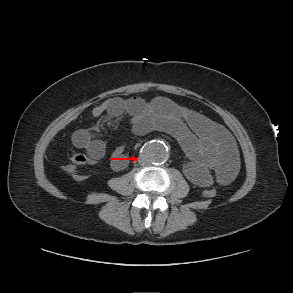
    • Infrarenal abdominal aortic aneurysm measuring 4.2 x 4 cm in cross-sectional diameter with discontinuous peripheral calcification and a focal outpouching along the right posterior wall
    • No retroperitoneal hematoma
    • Likely short segment dissection of the right common iliac artery with associated aneurysmal dilation measuring up to 2.5 cm in diameter
    • Moderate asymmetric atrophy of the right kidney
    • Cholecystectomy
    • Hysterectomy
  • MSK
    • No acute findings
    • Small fat-containing supraumbilical hernia and tiny fat-containing umbilical hernia


Diagnosis

  • Abdominal aortic aneurysm, impending rupture
  • Small bowel obstruction

Sample Report

Findings suggestive of high-grade small bowel obstruction with transition point in the right lower quadrant, likely relating to intraabdominal adhesions. A vascular etiology is difficult to exclude on this noncontrast study.

Infrarenal abdominal aortic aneurysm measuring up to 4.2 cm with discontinuous peripheral calcification and a focal outpouching along the right posterior wall. These findings are associated with an increased risk for aneurysm rupture, though there is no evidence for uncontained rupture at this time. Recommend evaluation by vascular surgery.

Focal dissection of the right common iliac artery, which is likely chronic but not well assessed on this noncontrast study.

Atrophic right kidney, which may relate to extensive atherosclerotic disease.

Left atrial and left ventricular dilation with findings suggestive of a chronic left ventricular apical infarct.

Mild pulmonary edema in the visualized lung bases.

Small hiatal hernia with fluid layering in the distal esophagus, which may relate to gastroesophageal reflux.


Discussion

Become a Pro Member to unlock the discussion.

View Plans   Log In


Images

Discontinuous mural calcification with a focal outpouching along the right posterior wall of an abdominal aortic aneurysm (red arrow), which places the patient at increased risk for future rupture.


📣 Feedback?

⌨️  Keyboard Shortcuts ("K")

Call Preparation

Prepare for call efficiently with interactive cases, sample reports, and annotated images.

1,400+ cases  91.25 CME

Call Simulator

   Neuro CT

250 cases

  Neuro MRI

146 cases

   Peds Radiographs

372 cases

   MSK Radiographs

281 cases

   Chest CT

84 cases

   Chest Radiographs

97 cases

   KUB

43 cases

   Body CT

140 cases

   Ultrasound

75 cases

Call Simulator

• Shuffle cases from our courses to simulate the mix of a call shift.
• Submit your own report before reviewing the case write-up.

Neuro Fellowship

Reviews of neuro topics with clinical pearls, differentials, and in-depth discussions.

131 cases  13.1 CME

   Brain Tumors

105 cases

   Neurodegenerative

13 cases

   Congenital Hearing Loss

13 cases

Call Preparation

   Neuro CT

250 cases

   Neuro MRI

146 cases

   Peds Radiographs

372 cases

   MSK Radiographs

281 cases

   Ultrasound

75 cases

   Chest CT

84 cases

   Chest Radiographs

97 cases

   Body CT

127 cases

   KUB

43 cases

Neuro Fellowship

   Brain Tumors

105 cases

   Neurodegenerative

13 cases

Call Simulator

• Shuffle cases from our courses.
• Submit your own report.

CME

• Earn up to 91 CME.
• Claim your CME to receive a certificate.

Call Simulator

• Shuffle cases from our courses to simulate the mix of a call shift.
• Submit your own report before reviewing the case write-up.

CME

• Earn up to 91 CME by completing cases in our radiology courses.
• Claim your CME to receive a certificate.

On Call

Quick references created specifically to help while on call.

  Peds Normals by Age

Reference database of normal imaging from birth to age 16

   Incidental Findings

Summary of consensus guidelines for managing incidental CT findings

   Media Index

Index of select illustrations & videos from our courses

  Neuro CT Mimics

Visual reference for common mimics of pathology on CT

On Call

  Peds Normals by Age

Reference database of normal imaging from birth to age 16

   Incidental Findings

Summary of consensus guidelines for managing incidental CT findings

   Media Index

Index of select illustrations & videos from our courses

  Neuro CT Mimics

Visual reference for common mimics of pathology on CT

Anatomy

Labelled radiographs and CT/MRI series teaching anatomy with a level of detail appropriate for medical students and junior residents.

   Pelvis

Pelvic MRI anatomy

   Chest

Chest radiograph & CT anatomy

   Body

Abdominal CT anatomy

   Cardiac

Cardiac CT anatomy

   Brain

Brain & calvarial anatomy on CT/MRI

   Cranial Nerves

Cranial nerves on MRI

   Shoulder

Shoulder MRI anatomy

   Knee

Knee MRI anatomy

   Temporal Bone

Resident/fellow-level anatomy

Anatomy

   Pelvis

Pelvic MRI anatomy

   Chest

Chest radiograph & CT anatomy

   Body

Abdominal CT anatomy

   Cardiac

Cardiac CT anatomy

   Brain

Brain & calvarial anatomy on CT/MRI

   Cranial Nerves

Cranial nerves on MRI

   Shoulder

Shoulder MRI anatomy

   Knee

Knee MRI anatomy

   Temporal Bone

Resident/fellow-level anatomy

Neuro Courses

Includes our call preparation neuro courses (Neuro CT and Neuro MRI) and our neuro fellowship courses.

   Neuro CT

250 cases

  Neuro MRI

146 cases

   Brain Tumors

105 cases

   Neurodegenerative

13 cases

   Congenital Hearing Loss

Coming soon

Neuro Courses

   Neuro CT

250 cases

   Neuro MRI

146 cases

   Brain Tumors

105 cases

   Neurodegenerative

13 cases

  Congenital Hearing Loss

Coming soon

CME

• Claim your CME to receive a certificate.

Call Simulator

• Shuffle cases from our courses to simulate the mix of a call shift.
• Submit your own report before reviewing the case write-up.

CME

• Earn up to 91 CME by completing cases in our radiology courses.
• Claim your CME to receive a certificate.

   Neuro CT

250 cases

   Neuro MRI

146 cases

   Peds Radiographs

372 cases

   MSK Radiographs

281 cases

   Ultrasound

75 cases

   Chest CT

84 cases

   Chest Radiographs

97 cases

   Body CT

140 cases

   KUB

43 cases

Call Simulator

• Shuffle cases from our courses to simulate the mix of a call shift.
• Submit your own report before reviewing the case write-up.

CME

• Earn up to 91 CME by completing cases in our radiology courses.
• Claim your CME to receive a certificate.

⌨️ Keyboard Shortcuts

View shortcuts

Zoom/Pan

  Full screen

  Window/Level

  Expand/collapse

  Scroll

  Save the case

  Close case/tab

Previous Slice (PC)

Next Slice (PC)

SHIFT Previous Slice (MAC)

 SHIFT Next Slice (MAC)

 Toggle presets

 Previous series (if multiple)

 Next series (if multiple)

  Previous case (⌘ ◄ on a mac)

  Next case (⌘ ► on a mac)

Toggle dark mode