Become a Pro Member for full access to this course and more.

View Plans   Log In
Expand ("E")   Collapse

Age: 36

Sex: Female

Indication: Left lower quadrant pain, shortness of breath

History: Right ovarian cancer status post en bloc resection of the mass including a portion of the right ureter, cytoreductive surgery, more recent left lower quadrant renal transplant

Save ("V")

Case #90


Findings

  • Chest
    • Mild enlargement of several left supraclavicular lymph nodes, with index nodes measuring up to 11 mm in short axis
    • No enlarged axillary, mediastinal, or hilar lymph nodes
    • Right IJ approach Port-A-Cath with catheter tip extending across the plane of the tricuspid valve into the right ventricle
    • Tunneled left IJ approach dialysis catheter with tip in the inferior aspect of the right atrium near the inferior cavoatrial junction
    • Mild dependent atelectasis/scarring
    • No suspicious pulmonary nodules
    • Small right pleural effusion
    • No pneumothorax
  • Abdomen/Pelvis
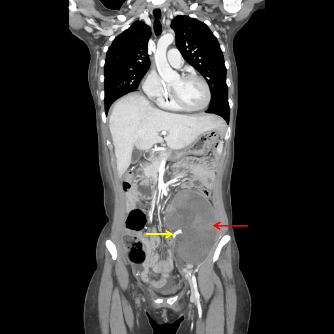
    • Enlarged left lower quadrant transplant kidney with diffuse hypoenhancement with the exception of a preserved rim of cortical enhancement
    • The transplant renal vein is not well visualized in the renal hilum, though the transplant renal artery and its proximal branches appear patent
    • Severe right hydronephrosis and proximal right hydroureter with an abrupt caliber change in the mid right ureter
    • Punctate nonobstructing right renal calculus
    • Surgical changes of retroperitoneal lymphadenectomy. No enlarged retroperitoneal lymph nodes
    • Mild presacral soft tissue thickening, likely treatment related
    • Small volume nonloculated ascites
    • Surgical changes of omentectomy
    • Surgical changes of hysterectomy and bilateral salpingo-oophorectomy
    • Heterogeneous enhancement of the liver parenchyma with multiple areas of wedge-shaped peripheral hypoattenuation
    • Mil diffuse pancreatic atrophy
    • Atherosclerotic calcification of the abdominal aorta and branch vessels, particularly affecting medium and small-sized arteries, without aneurysm
  • MSK
    • No acute osseous findings
    • Mild body wall edema
    • Ventral laparotomy changes


Diagnosis

  • Transplant renal vein thrombosis

Sample Report

Marked enlargement of the left lower quadrant transplant kidney with findings concerning for transplant renal vein thrombosis and developing renal infarction. Recommend urgent evaluation by transplant surgery.

Surgical changes following en bloc resection of a pelvic mass, cytoreductive debulking, and retroperitoneal lymphadenectomy. No findings specific for recurrent neoplasm in the abdomen or pelvis. Nonspecific mildly enlarged left supraclavicular lymph nodes which can be further assessed on followup imaging or PET/CT.

Severe right hydronephrosis and proximal hydroureter with abrupt caliber change in the mid right ureter, which may represent a postoperative stricture.

Heterogeneous hepatic parenchymal attenuation with multiple areas of wedge-shaped peripheral hypoattenuation, which may relate to hepatic congestion or altered hepatic perfusion, though hepatic infarcts could have a similar appearance. No findings highly concerning for hepatic metastatic disease.

Right IJ approach Port-A-Cath with catheter tip extending across the plane of the tricuspid valve into the right ventricle. Catheter repositioning should be considered.


Discussion

Become a Pro Member to unlock the discussion.

View Plans   Log In


Images

Enlarged, hypoenhancing left lower quadrant transplant kidney (red arrow) with visualization of the transplant artery (yellow arrow) but not the transplant vein in the renal hilum.


📣 Feedback?

⌨️  Keyboard Shortcuts ("K")

Call Preparation

Prepare for call efficiently with interactive cases, sample reports, and annotated images.

1,400+ cases  91.25 CME

Call Simulator

   Neuro CT

250 cases

  Neuro MRI

146 cases

   Peds Radiographs

372 cases

   MSK Radiographs

281 cases

   Chest CT

84 cases

   Chest Radiographs

97 cases

   KUB

43 cases

   Body CT

140 cases

   Ultrasound

75 cases

Call Simulator

• Shuffle cases from our courses to simulate the mix of a call shift.
• Submit your own report before reviewing the case write-up.

Neuro Fellowship

Reviews of neuro topics with clinical pearls, differentials, and in-depth discussions.

131 cases  13.1 CME

   Brain Tumors

105 cases

   Neurodegenerative

13 cases

   Congenital Hearing Loss

13 cases

Call Preparation

   Neuro CT

250 cases

   Neuro MRI

146 cases

   Peds Radiographs

372 cases

   MSK Radiographs

281 cases

   Ultrasound

75 cases

   Chest CT

84 cases

   Chest Radiographs

97 cases

   Body CT

127 cases

   KUB

43 cases

Neuro Fellowship

   Brain Tumors

105 cases

   Neurodegenerative

13 cases

Call Simulator

• Shuffle cases from our courses.
• Submit your own report.

CME

• Earn up to 91 CME.
• Claim your CME to receive a certificate.

Call Simulator

• Shuffle cases from our courses to simulate the mix of a call shift.
• Submit your own report before reviewing the case write-up.

CME

• Earn up to 91 CME by completing cases in our radiology courses.
• Claim your CME to receive a certificate.

On Call

Quick references created specifically to help while on call.

  Peds Normals by Age

Reference database of normal imaging from birth to age 16

   Incidental Findings

Summary of consensus guidelines for managing incidental CT findings

   Media Index

Index of select illustrations & videos from our courses

  Neuro CT Mimics

Visual reference for common mimics of pathology on CT

On Call

  Peds Normals by Age

Reference database of normal imaging from birth to age 16

   Incidental Findings

Summary of consensus guidelines for managing incidental CT findings

   Media Index

Index of select illustrations & videos from our courses

  Neuro CT Mimics

Visual reference for common mimics of pathology on CT

Anatomy

Labelled radiographs and CT/MRI series teaching anatomy with a level of detail appropriate for medical students and junior residents.

   Pelvis

Pelvic MRI anatomy

   Chest

Chest radiograph & CT anatomy

   Body

Abdominal CT anatomy

   Cardiac

Cardiac CT anatomy

   Brain

Brain & calvarial anatomy on CT/MRI

   Cranial Nerves

Cranial nerves on MRI

   Shoulder

Shoulder MRI anatomy

   Knee

Knee MRI anatomy

   Temporal Bone

Resident/fellow-level anatomy

Anatomy

   Pelvis

Pelvic MRI anatomy

   Chest

Chest radiograph & CT anatomy

   Body

Abdominal CT anatomy

   Cardiac

Cardiac CT anatomy

   Brain

Brain & calvarial anatomy on CT/MRI

   Cranial Nerves

Cranial nerves on MRI

   Shoulder

Shoulder MRI anatomy

   Knee

Knee MRI anatomy

   Temporal Bone

Resident/fellow-level anatomy

Neuro Courses

Includes our call preparation neuro courses (Neuro CT and Neuro MRI) and our neuro fellowship courses.

   Neuro CT

250 cases

  Neuro MRI

146 cases

   Brain Tumors

105 cases

   Neurodegenerative

13 cases

   Congenital Hearing Loss

Coming soon

Neuro Courses

   Neuro CT

250 cases

   Neuro MRI

146 cases

   Brain Tumors

105 cases

   Neurodegenerative

13 cases

  Congenital Hearing Loss

Coming soon

CME

• Claim your CME to receive a certificate.

Call Simulator

• Shuffle cases from our courses to simulate the mix of a call shift.
• Submit your own report before reviewing the case write-up.

CME

• Earn up to 91 CME by completing cases in our radiology courses.
• Claim your CME to receive a certificate.

   Neuro CT

250 cases

   Neuro MRI

146 cases

   Peds Radiographs

372 cases

   MSK Radiographs

281 cases

   Ultrasound

75 cases

   Chest CT

84 cases

   Chest Radiographs

97 cases

   Body CT

140 cases

   KUB

43 cases

Call Simulator

• Shuffle cases from our courses to simulate the mix of a call shift.
• Submit your own report before reviewing the case write-up.

CME

• Earn up to 91 CME by completing cases in our radiology courses.
• Claim your CME to receive a certificate.

⌨️ Keyboard Shortcuts

View shortcuts

Zoom/Pan

  Full screen

  Window/Level

  Expand/collapse

  Scroll

  Save the case

  Close case/tab

Previous Slice (PC)

Next Slice (PC)

SHIFT Previous Slice (MAC)

 SHIFT Next Slice (MAC)

 Toggle presets

 Previous series (if multiple)

 Next series (if multiple)

  Previous case (⌘ ◄ on a mac)

  Next case (⌘ ► on a mac)

Toggle dark mode