
Age: 53
Sex: Male
Indication: Trauma
Save ("V")
Case #31
Findings
Chest radiograph
- Mild widening of the superior mediastinum
- Endotracheal tube tip projects 4 cm above the carina
- Gastric suction catheter with side port overlying the GE junction and tip beyond the inferior margin of the radiograph
- Hazy airspace opacification in the right midlung with streaky bibasilar opacities
CT
- Chest
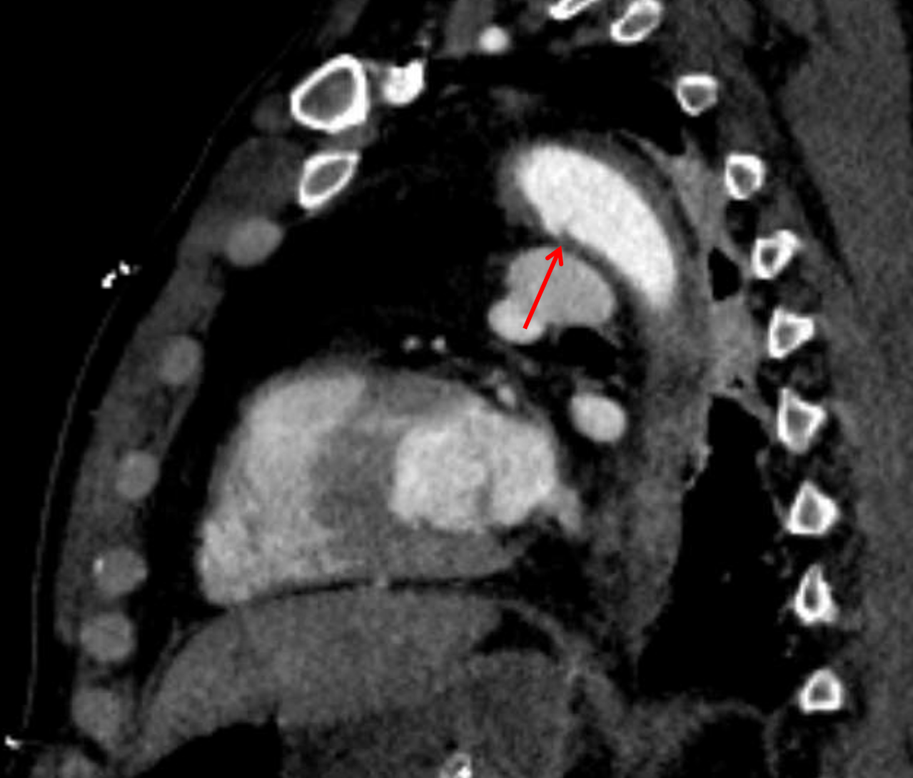
- Acute traumatic aortic intimal tear at the site of attachment of the ligamentum arteriosum with associated focal irregular bulge of the aortic external contour
- Small surrounding mediastinal hematoma
- Bilateral dependent airspace consolidation with patchy groundglass opacities in the right greater than left lungs
- Endotracheal tube terminates above the carina with a small amount of debris layering in the trachea
- Abdomen/pelvis
- Complex hepatic laceration in the right hepatic lobe predominantly involving segments 5 and 6 but extending into segments 7 and 8 with extension to the porta hepatis adjacent to the IVC. No evidence of active hemorrhage
- Small volume perihepatic hemoperitoneum tracking into the right paracolic gutter and into the anatomic pelvis
- Possible small laceration along the superior margin of the spleen measuring less than 1 cm with a small amount of perisplenic hemorrhage
- Right adrenal hematoma with hypoenhancement of the right adrenal gland
- Laceration of the inferomedial right kidney measuring 1.5 cm in depth with right subcapsular and perinephric hematoma. No evidence of active hemorrhage. No evidence of urine extravasation on delayed images
- Small amount of stranding along the intrahepatic IVC with a slit-like appearance of the IVC at the level of the renal veins
- Stranding adjacent to the second portion of the duodenum
- Gastric suction catheter terminates in the gastric body
- Urinary bladder is collapsed around a Foley catheter with intraluminal gas likely related to instrumentation
- MSK
- Acute minimally displaced right posterior first through seventh rib fractures
Diagnosis
- Acute traumatic aortic injury
- Hepatic laceration
- Splenic laceration
- Renal laceration
 Sample Report
Aortic intimal tear at the attachment of the ligamentum arteriosum with a small surrounding mediastinal hematoma and a small focal irregular bulge of the aortic external contour, which is consistent with a small pseudoaneurysm.
Acute minimally displaced right posterior first through seventh rib fractures with groundglass and consolidative pulmonary opacities likely represent a combination of contusion, aspiration, and atelectasis. No pneumothorax.
Complex hepatic laceration in the right hepatic lobe with extension to the porta hepatis adjacent to the IVC. Associated small volume perihepatic hemoperitoneum tracking into the right paracolic gutter and into the anatomic pelvis. Small amount of hemorrhage adjacent to the IVC could represent vascular injury, though there is no evidence of active hemorrhage.
Possible small laceration along the superior margin of the spleen measuring less than 1 cm with a small amount of perisplenic hemorrhage, though this is lower in attenuation than the perihepatic fluid.
Right adrenal hematoma with hypoenhancement of the right adrenal gland which may relate to contusion or vascular compromise.
Right renal laceration with subcapsular and perinephric hematoma. No evidence of urine extravasation on delayed images.
Slit-like appearance of the IVC at the level of the renal veins suggestive of hypovolemia.
Stranding adjacent to the second portion of the duodenum, which could relate to the adjacent hepatic and adrenal injuries, though a bowel injury is not excluded. Consider repeat imaging if there is high clinical concern for bowel injury.
Discussion

📣 Feedback?
⌨️ Keyboard Shortcuts ("K")
Help | Terms | Privacy Policy | Cookie Policy
Medical Disclaimer | © 2024 CaseStacks LLC
Related Cases
References
- Alonso RC, Nacenta SB, Martinez PD, Guerrero AS, Fuentes CG. Kidney in danger: CT findings of blunt and penetrating renal trauma. Radiographics 2009; 29(7): 2033-2053.
- Heneghan RE, Aarabi S, Quiroga E, Gunn ML, Singh N, Starnes BW. Call for a new classification system and treatment strategy in blunt aortic injury. J Vasc Surg 2016; 64(1): 171-176.
- . Adrenal gland hematomas in trauma patients. Radiology 2004; 230(3): 669-675.
- Saksobhavivat N, Shanmuganathan K, Chen HH, DuBose JJ, Richard H, Khan MA, Menaker J, Mirvis SE, Scalea TM. Blunt Splenic Injury: Use of a Multidetector CT-based Splenic Injury Grading System and Clinical Parameters for Triage of Patients at Admission. Radiology 2014; 3: 702-711.
- Soto JA, Anderson SW. Multidetector CT of blunt abdominal trauma. Radiology 2012; 265: 678-693.
- Steenburg S, Ravenel J, Ikonomidis J, Schonholz C, Reeves S. Acute traumatic aortic injury: Imaging evaluation and management. Radiology 2008; 248: 748-762.
- CT in blunt liver trauma. Radiographics 2005; 25(1): 87-104.

 View shortcuts
 Zoom/Pan
 Full screen
 Window/Level
 Expand/collapse
 Scroll
 Save the case
 Close case/tab


