Age: 54
Sex: Male
Indication: Pneumonia in immunocompromised patient
Save ("V")
Case #1
Findings
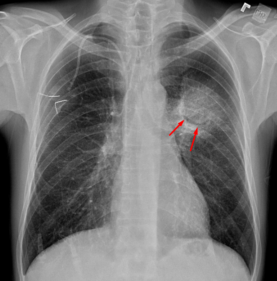
- Rounded opacity centered in the superior segment of the left lower lobe with a crescent of air noted. The opacity does not appear to displace or cross the major fissure
- No pleural effusion or pneumothorax
- Normal size and configuration of the cardiopericardial silhouette
Diagnosis
- Angioinvasive aspergillosis
 Sample Report
Rounded opacity centered in the superior segment of the left lower lobe with a crescent of air noted along its inferior aspect. This appearance is concerning for atypical pneumonia, in particular invasive aspergillosis. Consider chest CT for further evaluation.
Discussion
- Pulmonary aspergillosis has many different imaging appearances. Think about it as four different entities which are distinguished by the immune status of the patient:
- Hyperimmune – allergic bronchopulmonary aspergillosis (ABPA)
- Especially patients with asthma
- Upper lobe predominant bronchiectasis and bronchial plugging (finger-in-glove appearance)
- Normal immune – aspergilloma
- Patients with preexisting lung cavities
- Look for soft tissue density filling a cavity. A rim of gas around a fungal ball can look like a crescent, but is more correctly referred to as the Monod sign to separate this from the more ominous crescent sign of angioinvasive aspergillosis
- Mildly immunocompromised – semi-invasive (aka chronic necrotizing) aspergillosis
- Patients at risk include those with chronic diseases like diabetes, COPD, or malnutrition as well as those with chronic corticosteroid use
- Imaging features are very similar to those of reactivation tuberculosis with areas of consolidation and nodularity, sometimes with cavitation
- Full-blown immunocompromised – invasive aspergillosis
- Neutropenic patients and patients with AIDS
- Airways invasive subtype: bronchial thickening and tree-in-bud nodularity, which can appear identical to atypical bacterial infections
- Angioinvasive subtype: nodular opacities with groundglass halos or peripheral wedge shaped areas of consolidation. Air crescent sign can be seen during the healing phase as tissue necroses and retracts
 Images
📣 Feedback?
⌨️ Keyboard Shortcuts ("K")
Help | Terms | Privacy Policy | Cookie Policy
Medical Disclaimer | © 2024 CaseStacks LLC
Related Cases
Temporarily disabled.

 View shortcuts
 Zoom/Pan
 Full screen
 Window/Level
 Expand/collapse
 Scroll
 Save the case
 Close case/tab


