Purchase the medical student plan for full access to this course.

View Pricing   Log In
Expand ("E")   Collapse

Age: 67

Sex: Female

Indication: Chest pain, stroke-like symptoms

Save ("V")

Case #4


Findings

  • Vascular findings
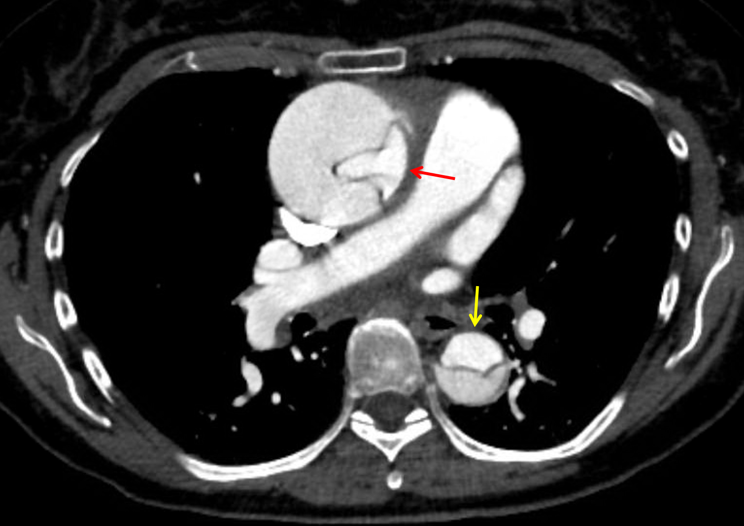
    • Stanford type A aortic dissection extending from the aortic annulus at the level of the noncoronary aortic valve leaflet throughout the entire aorta and into the left common iliac artery
    • Resultant aneurysmal dilation of the ascending aorta measuring up to 5.9 x 5.7 cm in cross-sectional diameter
    • Moderate compression of the true lumen, which remains patent
    • Dissection does not extend into the right or left main coronary arteries, though the false lumen does exert mass effect on the right and left coronary cusps
    • Dissection extends into the brachiocephalic artery with resulting severe narrowing of the true lumen as well as occlusion of the origin of the right common carotid artery and severe narrowing at the origin of the right subclavian artery
    • Dissection extends slightly into the origins of the left common carotid and left subclavian arteries without high-grade stenosis
    • Linear intraluminal hypodensity in the proximal left common carotid artery
    • Celiac trunk primarily arises from the true lumen with some contribution from the false lumen
    • SMA, renal arteries, and IMA arise from the true lumen and are patent
    • It is unclear whether the left internal iliac artery arises from the true or false lumen
    • No hemopericardium or hemothorax
  • Nonvascular findings
    • Mild cardiomegaly
    • Small hiatal hernia
    • Mild centrilobular emphysema
    • Mild widespread bronchial wall thickening
    • Ill-defined area of groundglass opacification in the lingula
    • Mild bilateral subpleural reticulation, suggesting mild fibrosis
    • Scattered hypodense liver lesions, many of which are too small to characterize
    • Cholelithiasis
    • 2.7 cm enhancing solid mass arising from the upper pole of the right kidney
    • Colonic diverticulosis


Diagnosis

  • Aortic dissection

Sample Report

Stanford type A aortic dissection originating at the noncoronary cusp of the aortic annulus, extends through the thoracic and abdominal aorta, and terminates in the left common iliac artery. Resultant aneurysmal dilation of the ascending aorta measuring up to 5.9 cm. The coronary arteries arise from the true lumen and are patent. No hemopericardium or hemothorax.

Dissection extends into the arch vessels with notable occlusion of the proximal right common carotid artery and severe narrowing of the brachiocephalic artery and right subclavian artery.

A linear intraluminal hypodensity in the proximal left common carotid artery may be artifactual, though a focal dissection at this location is not excluded. Recommend attention on followup imaging.

Mesenteric vessels arise from the true lumen and are patent, though there is a minor contribution to the celiac artery from the false lumen.

Enhancing solid right upper pole renal mass concerning for renal cell carcinoma.

Ill-defined area of groundglass opacification in the lingula, which may be infectious or inflammatory in etiology. Recommend attention on followup imaging to exclude malignancy.


Discussion


Purchase the medical student plan for full access to this course.

View Pricing   Log In


Images

Stanford type A aortic dissection with the true lumen compressed medially in the ascending aorta (red arrow) and anteriorly in the descending aorta (yellow arrow).


📣 Feedback?

⌨️  Keyboard Shortcuts ("K")

Call Preparation

Prepare for call efficiently with interactive cases, sample reports, and annotated images.

1,400+ cases  91.25 CME

Call Simulator

   Neuro CT

250 cases

  Neuro MRI

146 cases

   Peds Radiographs

372 cases

   MSK Radiographs

281 cases

   Chest CT

84 cases

   Chest Radiographs

97 cases

   KUB

43 cases

   Body CT

140 cases

   Ultrasound

75 cases

Call Simulator

• Shuffle cases from our courses to simulate the mix of a call shift.
• Submit your own report before reviewing the case write-up.

Neuro Fellowship

Reviews of neuro topics with clinical pearls, differentials, and in-depth discussions.

131 cases  13.1 CME

   Brain Tumors

105 cases

   Neurodegenerative

13 cases

   Congenital Hearing Loss

13 cases

Call Preparation

   Neuro CT

250 cases

   Neuro MRI

146 cases

   Peds Radiographs

372 cases

   MSK Radiographs

281 cases

   Ultrasound

75 cases

   Chest CT

84 cases

   Chest Radiographs

97 cases

   Body CT

127 cases

   KUB

43 cases

Neuro Fellowship

   Brain Tumors

105 cases

   Neurodegenerative

13 cases

Call Simulator

• Shuffle cases from our courses.
• Submit your own report.

CME

• Earn up to 91 CME.
• Claim your CME to receive a certificate.

Call Simulator

• Shuffle cases from our courses to simulate the mix of a call shift.
• Submit your own report before reviewing the case write-up.

CME

• Earn up to 91 CME by completing cases in our radiology courses.
• Claim your CME to receive a certificate.

On Call

Quick references created specifically to help while on call.

  Peds Normals by Age

Reference database of normal imaging from birth to age 16

   Incidental Findings

Summary of consensus guidelines for managing incidental CT findings

   Media Index

Index of select illustrations & videos from our courses

  Neuro CT Mimics

Visual reference for common mimics of pathology on CT

On Call

  Peds Normals by Age

Reference database of normal imaging from birth to age 16

   Incidental Findings

Summary of consensus guidelines for managing incidental CT findings

   Media Index

Index of select illustrations & videos from our courses

  Neuro CT Mimics

Visual reference for common mimics of pathology on CT

Anatomy

Labelled radiographs and CT/MRI series teaching anatomy with a level of detail appropriate for medical students and junior residents.

   Pelvis

Pelvic MRI anatomy

   Chest

Chest radiograph & CT anatomy

   Body

Abdominal CT anatomy

   Cardiac

Cardiac CT anatomy

   Brain

Brain & calvarial anatomy on CT/MRI

   Cranial Nerves

Cranial nerves on MRI

   Shoulder

Shoulder MRI anatomy

   Knee

Knee MRI anatomy

   Temporal Bone

Resident/fellow-level anatomy

Anatomy

   Pelvis

Pelvic MRI anatomy

   Chest

Chest radiograph & CT anatomy

   Body

Abdominal CT anatomy

   Cardiac

Cardiac CT anatomy

   Brain

Brain & calvarial anatomy on CT/MRI

   Cranial Nerves

Cranial nerves on MRI

   Shoulder

Shoulder MRI anatomy

   Knee

Knee MRI anatomy

   Temporal Bone

Resident/fellow-level anatomy

Neuro Courses

Includes our call preparation neuro courses (Neuro CT and Neuro MRI) and our neuro fellowship courses.

   Neuro CT

250 cases

  Neuro MRI

146 cases

   Brain Tumors

105 cases

   Neurodegenerative

13 cases

   Congenital Hearing Loss

Coming soon

Neuro Courses

   Neuro CT

250 cases

   Neuro MRI

146 cases

   Brain Tumors

105 cases

   Neurodegenerative

13 cases

  Congenital Hearing Loss

Coming soon

CME

• Claim your CME to receive a certificate.

Call Simulator

• Shuffle cases from our courses to simulate the mix of a call shift.
• Submit your own report before reviewing the case write-up.

CME

• Earn up to 91 CME by completing cases in our radiology courses.
• Claim your CME to receive a certificate.

   Neuro CT

250 cases

   Neuro MRI

146 cases

   Peds Radiographs

372 cases

   MSK Radiographs

281 cases

   Ultrasound

75 cases

   Chest CT

84 cases

   Chest Radiographs

97 cases

   Body CT

140 cases

   KUB

43 cases

Call Simulator

• Shuffle cases from our courses to simulate the mix of a call shift.
• Submit your own report before reviewing the case write-up.

CME

• Earn up to 91 CME by completing cases in our radiology courses.
• Claim your CME to receive a certificate.

⌨️ Keyboard Shortcuts

View shortcuts

Zoom/Pan

  Full screen

  Window/Level

  Expand/collapse

  Scroll

  Save the case

  Close case/tab

Previous Slice (PC)

Next Slice (PC)

SHIFT Previous Slice (MAC)

 SHIFT Next Slice (MAC)

 Toggle presets

 Previous series (if multiple)

 Next series (if multiple)

  Previous case (⌘ ◄ on a mac)

  Next case (⌘ ► on a mac)

Toggle dark mode