Become a Pro Member for full access to this course and more.

View Plans   Log In
Expand ("E")   Collapse

Age: 33

Sex: Male

Indication: Trauma

Save ("V")

Case #1


Findings

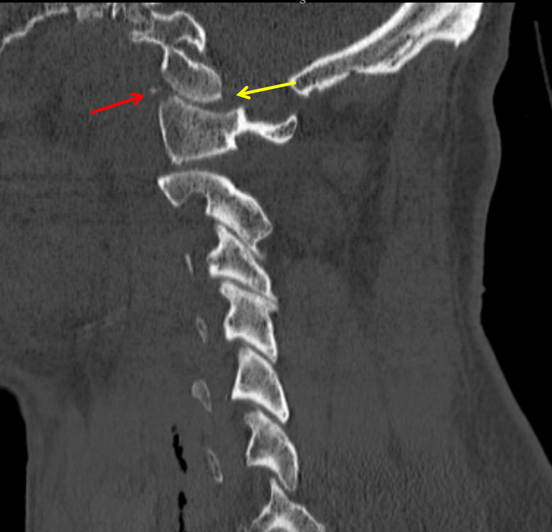
  • Multiple tiny acute displaced fractures along the anterior aspects of both C1 superior articular facets with mild anterior subluxation of the occipital condyles relative to C1 and widening of the posterior aspect of both atlanto-occipital joints
  • Widening of the C1-C2 interspinous interval and of the superior aspect of the sagittal atlantodental interval
  • Extensive prevertebral edema extending from the skull base through the C5 level
  • Comminuted, displaced acute left posterior first through fourth rib fractures
  • Left chest tube terminates in the medial left apex, passing immediately posterior to the left subclavian artery and contacting the left lateral aspect of the trachea.
  • Tiny left pneumothorax. Extensive left apical pulmonary contusion with small layering hemothorax.
  • Deep soft tissue emphysema in the base of the neck


Diagnosis

  • Fracture-subluxation at the craniocervical junction

Sample Report

Acute fracture-subluxation at the craniocervical junction with multiple tiny acute displaced fractures along the anterior aspects of both C1 superior articular facets, mild anterior subluxation of the occipital condyles relative to C1, widening of the posterior aspect of both atlanto-occipital joints, widening of the C1-C2 interspinous interval, and widening of the superior aspect of the sagittal atlantodental interval. MRI could further evaluate the extent of ligamentous injury.

Extensive upper and mid cervical prevertebral edema.

Comminuted, displaced left first through fourth rib fractures with left apical pulmonary contusion and a small left hemopneumothorax. Left chest tube terminates in the medial left apex, passing immediately posterior to the left subclavian artery and contacting the left lateral aspect of the trachea.


Discussion

Become a Pro Member to unlock the discussion.

View Plans   Log In


Images

Red arrow: displaced fracture fragments along the anterior aspect of the C1 superior articular facets. Yellow arrow: anterior subluxation of the occipital condyle relative to C1 with widening of the posterior aspect of the atlanto-occipital joint. Postoperative radiograph in this patient. Sagittal view of the major cervical spine ligaments. Coronal view of the major ligaments at the craniocervical junction.


📣 Feedback?

⌨️  Keyboard Shortcuts ("K")

Call Preparation

Prepare for call efficiently with interactive cases, sample reports, and annotated images.

1,400+ cases  91.25 CME

Call Simulator

   Neuro CT

250 cases

  Neuro MRI

146 cases

   Peds Radiographs

372 cases

   MSK Radiographs

281 cases

   Chest CT

84 cases

   Chest Radiographs

97 cases

   KUB

43 cases

   Body CT

140 cases

   Ultrasound

75 cases

Call Simulator

• Shuffle cases from our courses to simulate the mix of a call shift.
• Submit your own report before reviewing the case write-up.

Neuro Fellowship

Reviews of neuro topics with clinical pearls, differentials, and in-depth discussions.

131 cases  13.1 CME

   Brain Tumors

105 cases

   Neurodegenerative

13 cases

   Congenital Hearing Loss

13 cases

Call Preparation

   Neuro CT

250 cases

   Neuro MRI

146 cases

   Peds Radiographs

372 cases

   MSK Radiographs

281 cases

   Ultrasound

75 cases

   Chest CT

84 cases

   Chest Radiographs

97 cases

   Body CT

127 cases

   KUB

43 cases

Neuro Fellowship

   Brain Tumors

105 cases

   Neurodegenerative

13 cases

Call Simulator

• Shuffle cases from our courses.
• Submit your own report.

CME

• Earn up to 91 CME.
• Claim your CME to receive a certificate.

Call Simulator

• Shuffle cases from our courses to simulate the mix of a call shift.
• Submit your own report before reviewing the case write-up.

CME

• Earn up to 91 CME by completing cases in our radiology courses.
• Claim your CME to receive a certificate.

On Call

Quick references created specifically to help while on call.

  Peds Normals by Age

Reference database of normal imaging from birth to age 16

   Incidental Findings

Summary of consensus guidelines for managing incidental CT findings

   Media Index

Index of select illustrations & videos from our courses

  Neuro CT Mimics

Visual reference for common mimics of pathology on CT

On Call

  Peds Normals by Age

Reference database of normal imaging from birth to age 16

   Incidental Findings

Summary of consensus guidelines for managing incidental CT findings

   Media Index

Index of select illustrations & videos from our courses

  Neuro CT Mimics

Visual reference for common mimics of pathology on CT

Anatomy

Labelled radiographs and CT/MRI series teaching anatomy with a level of detail appropriate for medical students and junior residents.

   Pelvis

Pelvic MRI anatomy

   Chest

Chest radiograph & CT anatomy

   Body

Abdominal CT anatomy

   Cardiac

Cardiac CT anatomy

   Brain

Brain & calvarial anatomy on CT/MRI

   Cranial Nerves

Cranial nerves on MRI

   Shoulder

Shoulder MRI anatomy

   Knee

Knee MRI anatomy

   Temporal Bone

Resident/fellow-level anatomy

Anatomy

   Pelvis

Pelvic MRI anatomy

   Chest

Chest radiograph & CT anatomy

   Body

Abdominal CT anatomy

   Cardiac

Cardiac CT anatomy

   Brain

Brain & calvarial anatomy on CT/MRI

   Cranial Nerves

Cranial nerves on MRI

   Shoulder

Shoulder MRI anatomy

   Knee

Knee MRI anatomy

   Temporal Bone

Resident/fellow-level anatomy

Neuro Courses

Includes our call preparation neuro courses (Neuro CT and Neuro MRI) and our neuro fellowship courses.

   Neuro CT

250 cases

  Neuro MRI

146 cases

   Brain Tumors

105 cases

   Neurodegenerative

13 cases

   Congenital Hearing Loss

Coming soon

Neuro Courses

   Neuro CT

250 cases

   Neuro MRI

146 cases

   Brain Tumors

105 cases

   Neurodegenerative

13 cases

  Congenital Hearing Loss

Coming soon

CME

• Claim your CME to receive a certificate.

Call Simulator

• Shuffle cases from our courses to simulate the mix of a call shift.
• Submit your own report before reviewing the case write-up.

CME

• Earn up to 91 CME by completing cases in our radiology courses.
• Claim your CME to receive a certificate.

   Neuro CT

250 cases

   Neuro MRI

146 cases

   Peds Radiographs

372 cases

   MSK Radiographs

281 cases

   Ultrasound

75 cases

   Chest CT

84 cases

   Chest Radiographs

97 cases

   Body CT

140 cases

   KUB

43 cases

Call Simulator

• Shuffle cases from our courses to simulate the mix of a call shift.
• Submit your own report before reviewing the case write-up.

CME

• Earn up to 91 CME by completing cases in our radiology courses.
• Claim your CME to receive a certificate.

⌨️ Keyboard Shortcuts

View shortcuts

Zoom/Pan

  Full screen

  Window/Level

  Expand/collapse

  Scroll

  Save the case

  Close case/tab

Previous Slice (PC)

Next Slice (PC)

SHIFT Previous Slice (MAC)

 SHIFT Next Slice (MAC)

 Toggle presets

 Previous series (if multiple)

 Next series (if multiple)

  Previous case (⌘ ◄ on a mac)

  Next case (⌘ ► on a mac)

Toggle dark mode