
Age: 55
Sex: Male
Indication: Trauma
Save ("V")
Case #5
Findings
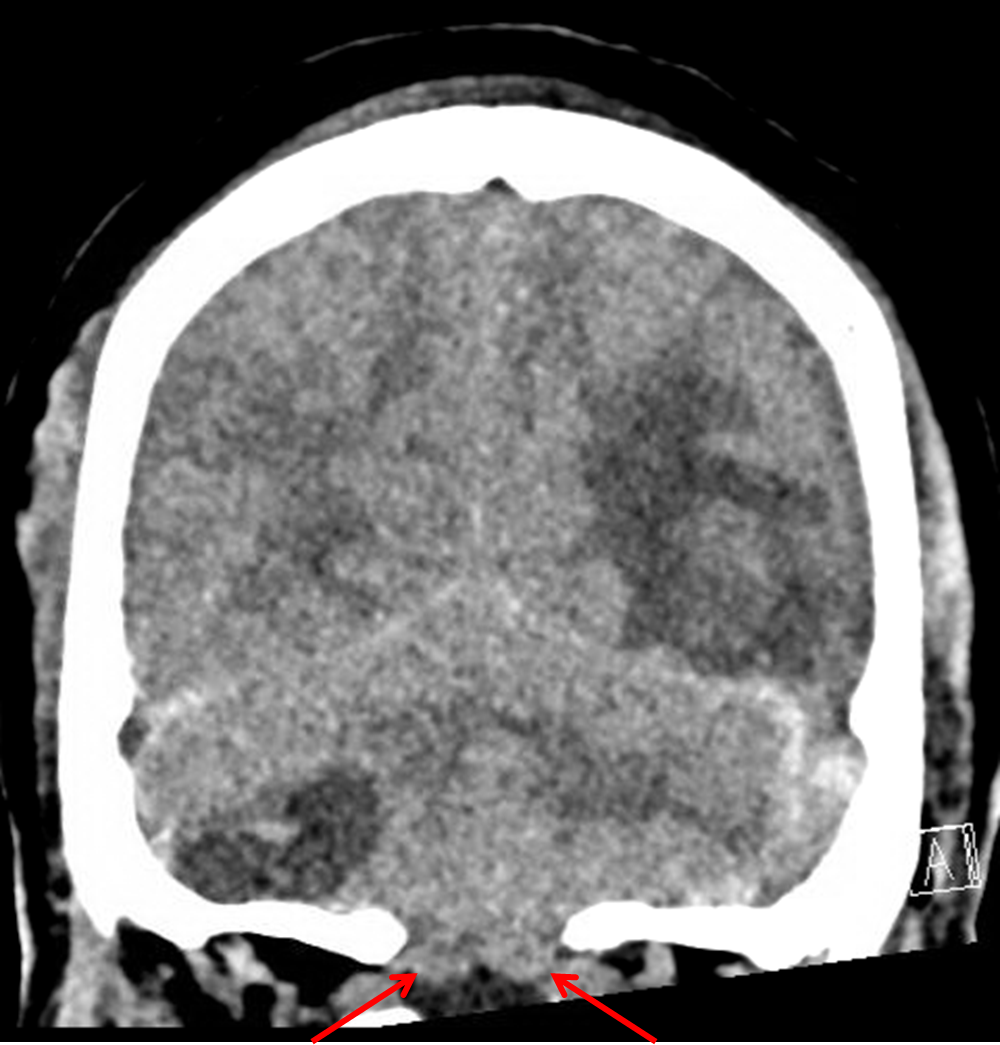
- Acute nondepressed occipital bone fracture extending into the left lambdoid suture with widening of the left occipitomastoid suture and possible extension into the mastoid temporal bone
- Acute left temporo-parieto-occipital intraparenchymal hematoma with surrounding vasogenic edema
- Small volume acute subdural hemorrhage layering over the bilateral cerebral convexities, along the interhemispheric falx, and along the bilateral cerebellar tentorial leaflets
- Mixed density right greater than left extra-axial infratentorial collections with layering low density on the right
- Cerebellar tonsillar and upward transtentorial herniation with crowding of the foramen magnum and effacement of the fourth ventricle and basal cisterns
- Trace blood products layering in the occipital horn of the right lateral ventricle
- Left frontal approach ventriculostomy catheter traverses the foramen of Monro terminating in the region of the suprasellar cistern with low density along the catheter track. Slight enlargement of the temporal horns of the lateral ventricles
- Multifocal scalp contusion with skin staples along the high posterior scalp
- Partial opacification of bilateral mastoid air cells without definite temporal bone fracture
- Inflammatory mucosal thickening of the paranasal sinuses
- Partially imaged nasoenteric tube
Diagnosis
- Multicompartmental hemorrhage with tonsillar herniation
 Sample Report
Acute nondepressed occipital bone fracture extending into the left lambdoid suture with widening of the left occipitomastoid suture and possible extension into the mastoid temporal bone.
Acute multicompartmental intracranial hemorrhage with the following components: left temporo-parieto-occipital intraparenchymal hematoma with surrounding vasogenic edema, small volume acute subdural hemorrhage layering over the bilateral cerebral convexities (measuring x mm on the right and x mm on the left), along the interhemispheric falx, and along the bilateral cerebellar tentorial leaflets, trace blood products layering in the occipital horn of the right lateral ventricle, and right greater than left extra-axial infratentorial hematomas. Layering low density in the right posterior fossa collection is concerning for unclotted blood which may relate to hyperacute hemorrhage or coagulopathy.
Extensive posterior fossa mass effect resulting in cerebellar tonsillar and upward transtentorial herniation with crowding of the foramen magnum and effacement of the fourth ventricle and basal cisterns.
Left frontal approach ventriculostomy catheter traverses the foramen of Monro terminating in the region of the suprasellar cistern with edema/developing gliosis along the catheter track. Slight enlargement of the temporal horns of the lateral ventricles.
Partial opacification of bilateral mastoid air cells without definite temporal bone fracture.
Multifocal scalp contusion with skin staples along the high posterior scalp.


 View shortcuts
 Zoom/Pan
 Full screen
 Window/Level
 Expand/collapse
 Scroll
 Save the case
 Close case/tab


