Expand ("E")   Collapse

Age: 91

Sex: Female

Indication: Left-sided weakness

Save ("V")

Case #1


Findings

  • Large area of restricted diffusion in the right MCA territory
  • Associated T2/FLAIR hyperintensity and swelling with right cerebral sulcal effacement and 3 mm right-to-left midline shift
  • Basal cisterns are patent
  • Abnormal flow-related signal in the communicating segment of the right internal carotid artery and in the M1 segment of the right MCA
  • Few small foci of susceptibility artifact in the right frontal lobe
  • Confluent subcortical and periventricular white matter T2/FLAIR hyperintensity
  • Remote lacunar infarcts in the pons
  • Generalized cerebral volume loss
  • Bilateral pseudophakia


Diagnosis

  • Thrombotic stroke

Sample Report

Large acute/early subacute right MCA territory infarct. Associated cytotoxic edema with resultant right cerebral sulcal effacement and 3 mm right-to-left midline shift. No evidence of herniation or hydrocephalus.

Few small foci of susceptibility artifact in the right frontal lobe may represent petechial hemorrhage, though there is no evidence of organized hemorrhagic conversion.

Abnormal flow-related signal in the communicating segment of the right internal carotid artery and in the M1 segment of the right MCA concerning for diminished/slow flow or occlusion. Recommend correlation with CTA or catheter angiography.

Confluent subcortical and periventricular white matter T2/FLAIR hyperintensity, which though nonspecific likely relates to chronic small vessel disease. Remote lacunar infarcts in the pons. Generalized cerebral volume loss.


Discussion

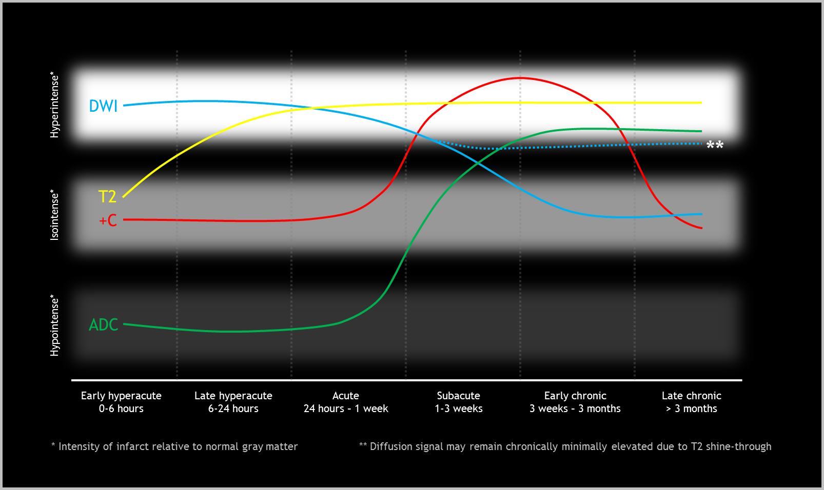
  • MRI is much more sensitive than CT for the detection of early cerebral infarcts
  • Abnormal signal first becomes apparent on diffusion/ADC, often present by mere minutes after the vascular occlusion occurs
    • ADC signal normalizes more quickly (typically by 10 days) than diffusion signal because of T2 effects which may transiently or permanently keep diffusion signal mildly elevated
  • T2/FLAIR signal abnormality lags somewhat, typically starting to become apparent 6-8 hours after the occlusion
  • There are three types of enhancement that can be observed with strokes:
    1. Arterial enhancement (hours to days after stroke) – decreased arterial blood flow within the area of acute ischemia results in decreased flow-related signal loss. This increased intrinsic signal combined with the signal from administered contrast material makes these vessels appear more hyperintense than unaffected arteries
    2. Meningeal enhancement (1-7 days after stroke) – resulting from reactive hyperemia in the meninges overlying the infarct. This is the least commonly observed type of enhancement
    3. Parenchymal enhancement (1 week – 3 months) – resulting from breakdown of the blood-brain barrier. Parenchymal enhancement may occur earlier and more intensely in incomplete infarcts. If you are trying to determine if an enhancing lesion is an infarct vs neoplasm, repeating imaging in a few months can help differentiate the two
  • Refer to the figure below for a graphical representation of the typical progression of MR signal in cerebral vascular accidents


Images

Evolution of signal on several MRI sequences in cerebral infarcts. +C = postcontrast imaging.


📣 Feedback?

⌨️  Keyboard Shortcuts ("K")

Call Preparation

Prepare for call efficiently with interactive cases, sample reports, and annotated images.

1,400+ cases  91.25 CME

Call Simulator

   Neuro CT

250 cases

  Neuro MRI

146 cases

   Peds Radiographs

372 cases

   MSK Radiographs

281 cases

   Chest CT

84 cases

   Chest Radiographs

97 cases

   KUB

43 cases

   Body CT

140 cases

   Ultrasound

75 cases

Call Simulator

• Shuffle cases from our courses to simulate the mix of a call shift.
• Submit your own report before reviewing the case write-up.

Neuro Fellowship

Reviews of neuro topics with clinical pearls, differentials, and in-depth discussions.

131 cases  13.1 CME

   Brain Tumors

105 cases

   Neurodegenerative

13 cases

   Congenital Hearing Loss

13 cases

Call Preparation

   Neuro CT

250 cases

   Neuro MRI

146 cases

   Peds Radiographs

372 cases

   MSK Radiographs

281 cases

   Ultrasound

75 cases

   Chest CT

84 cases

   Chest Radiographs

97 cases

   Body CT

127 cases

   KUB

43 cases

Neuro Fellowship

   Brain Tumors

105 cases

   Neurodegenerative

13 cases

Call Simulator

• Shuffle cases from our courses.
• Submit your own report.

CME

• Earn up to 91 CME.
• Claim your CME to receive a certificate.

Call Simulator

• Shuffle cases from our courses to simulate the mix of a call shift.
• Submit your own report before reviewing the case write-up.

CME

• Earn up to 91 CME by completing cases in our radiology courses.
• Claim your CME to receive a certificate.

On Call

Quick references created specifically to help while on call.

  Peds Normals by Age

Reference database of normal imaging from birth to age 16

   Incidental Findings

Summary of consensus guidelines for managing incidental CT findings

   Media Index

Index of select illustrations & videos from our courses

  Neuro CT Mimics

Visual reference for common mimics of pathology on CT

On Call

  Peds Normals by Age

Reference database of normal imaging from birth to age 16

   Incidental Findings

Summary of consensus guidelines for managing incidental CT findings

   Media Index

Index of select illustrations & videos from our courses

  Neuro CT Mimics

Visual reference for common mimics of pathology on CT

Anatomy

Labelled radiographs and CT/MRI series teaching anatomy with a level of detail appropriate for medical students and junior residents.

   Pelvis

Pelvic MRI anatomy

   Chest

Chest radiograph & CT anatomy

   Body

Abdominal CT anatomy

   Cardiac

Cardiac CT anatomy

   Brain

Brain & calvarial anatomy on CT/MRI

   Cranial Nerves

Cranial nerves on MRI

   Shoulder

Shoulder MRI anatomy

   Knee

Knee MRI anatomy

   Temporal Bone

Resident/fellow-level anatomy

Anatomy

   Pelvis

Pelvic MRI anatomy

   Chest

Chest radiograph & CT anatomy

   Body

Abdominal CT anatomy

   Cardiac

Cardiac CT anatomy

   Brain

Brain & calvarial anatomy on CT/MRI

   Cranial Nerves

Cranial nerves on MRI

   Shoulder

Shoulder MRI anatomy

   Knee

Knee MRI anatomy

   Temporal Bone

Resident/fellow-level anatomy

Neuro Courses

Includes our call preparation neuro courses (Neuro CT and Neuro MRI) and our neuro fellowship courses.

   Neuro CT

250 cases

  Neuro MRI

146 cases

   Brain Tumors

105 cases

   Neurodegenerative

13 cases

   Congenital Hearing Loss

Coming soon

Neuro Courses

   Neuro CT

250 cases

   Neuro MRI

146 cases

   Brain Tumors

105 cases

   Neurodegenerative

13 cases

  Congenital Hearing Loss

Coming soon

CME

• Claim your CME to receive a certificate.

Call Simulator

• Shuffle cases from our courses to simulate the mix of a call shift.
• Submit your own report before reviewing the case write-up.

CME

• Earn up to 91 CME by completing cases in our radiology courses.
• Claim your CME to receive a certificate.

   Neuro CT

250 cases

   Neuro MRI

146 cases

   Peds Radiographs

372 cases

   MSK Radiographs

281 cases

   Ultrasound

75 cases

   Chest CT

84 cases

   Chest Radiographs

97 cases

   Body CT

140 cases

   KUB

43 cases

Call Simulator

• Shuffle cases from our courses to simulate the mix of a call shift.
• Submit your own report before reviewing the case write-up.

CME

• Earn up to 91 CME by completing cases in our radiology courses.
• Claim your CME to receive a certificate.

⌨️ Keyboard Shortcuts

View shortcuts

Zoom/Pan

  Full screen

  Window/Level

  Expand/collapse

  Scroll

  Save the case

  Close case/tab

Previous Slice (PC)

Next Slice (PC)

SHIFT Previous Slice (MAC)

 SHIFT Next Slice (MAC)

 Toggle presets

 Previous series (if multiple)

 Next series (if multiple)

  Previous case (⌘ ◄ on a mac)

  Next case (⌘ ► on a mac)

Toggle dark mode