Become a Pro Member for full access to this course and more.

View Plans   Log In
Expand ("E")   Collapse

Age: 53

Sex: Female

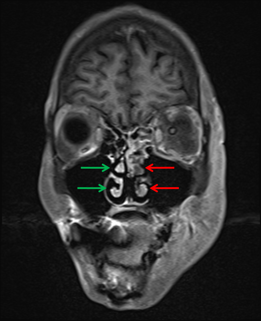
Indication: Left eye blindness

Save ("V")

Case #37


Findings

  • T2/FLAIR signal hyperintensity in the left sphenoid sinus, left greater than right maxillary sinuses, and throughout bilateral ethmoid air cells
  • Left periorbital and postseptal T2/FLAIR signal hyperintensity and enhancement with slight left proptosis
  • T2/FLAIR signal hyperintensity and enhancement in the left masticator space involving the left muscles of mastication and extending into the left parapharyngeal space
  • No peripherally enhancing collection
  • Restricted diffusion involving the orbital and canalicular segments of the left optic nerve
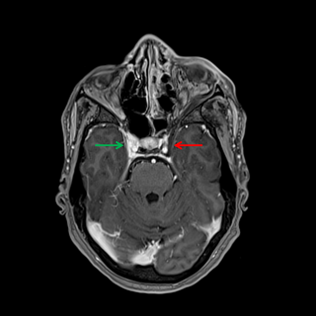
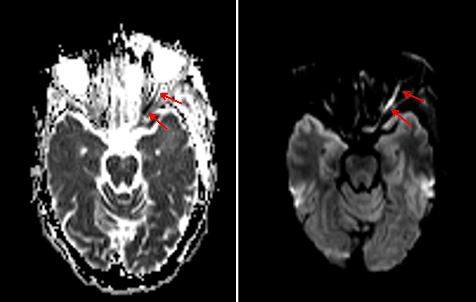
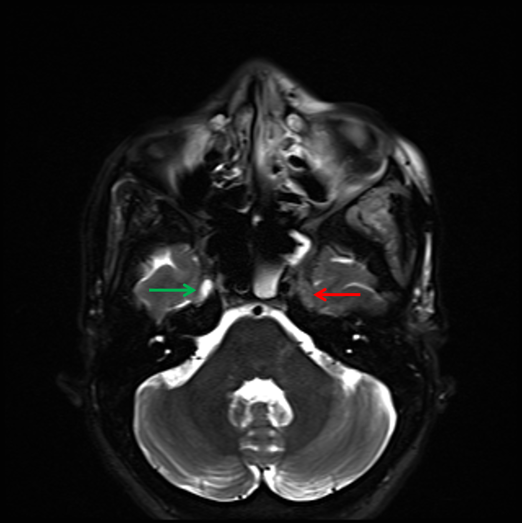
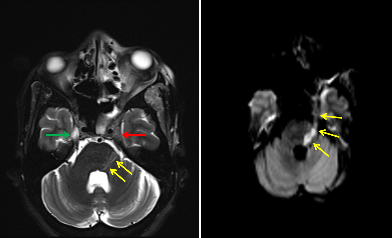
  • Abnormal T2 hypointensity in the left foramen ovale and left Meckel’s cave with restricted diffusion involving the cisternal segment of the left trigeminal nerve extending into the left dorsolateral pons and left Meckel’s cave
  • Asymmetric hypoenhancement of the left superior ophthalmic vein and left cavernous sinus
  • No convincing contrast opacification of the left ophthalmic artery, though the ophthalmic segment of the left internal carotid artery opacifies
  • Hypoenhancement of the left middle and inferior nasal turbinates


Diagnosis

  • Cavernous sinus thrombosis
  • Invasive mucormycosis

Sample Report

Findings highly concerning for invasive fungal sinusitis with sinus disease involving the left sphenoid sinus, left greater than right maxillary sinuses, and bilateral ethmoid air cells and contiguous extension with left preseptal and postseptal orbital cellulitis, cellulitis/myositis involving the left masticator and parapharyngeal spaces, and evidence of intracranial extension with left superior ophthalmic vein and left cavernous sinus thrombosis. No peripherally enhancing collection to suggest abscess. No convincing enhancement of the left ophthalmic artery is concerning for thrombosis, which could be further evaluated with CTA or catheter angiography. Recommend urgent surgical consultation.

Restricted diffusion involving the orbital and canalicular segments of the left optic nerve concerning for infectious involvement and/or infarct.

Findings consistent with perineural spread of infection along the V3 branch of the left trigeminal nerve from the left masticator space, through foramen ovale, and involving the left Meckel’s cave with further retrograde extension along the cisternal segment of the left trigeminal nerve into the left dorsolateral pons.

Hypoenhancement of the left middle and inferior nasal turbinates concerning for necrosis.


Discussion

Become a Pro Member to unlock the discussion.

View Plans   Log In


Images

Abnormal nonopacification of the left cavernous sinus (red arrow) compared to the normal right side (green arrow) indicative of cavernous sinus thrombosis. Restricted diffusion involving the orbital and canalicular segments of the left optic nerve (red arrows) concerning for infectious involvement and/or ischemia. Abnormal T2 signal hypointensity in the left Meckel's cave (red arrow) compared to the normal appearance on the right (green arrow) concerning for infectious involvement. Abnormal T2 signal hypointensity in the left Meckel's cave (red arrow) compared to the normal appearance on the right (green arrow) with restricted diffusion extending from the left Meckel's cave along the cisternal segment of the left trigeminal nerve into the dorsolateral pons (yellow arrows) concerning for perineural spread of infection to the brainstem. Hypoenhancement of the left middle and inferior nasal turbinates (red arrows) compared to the normal appearance on the right (green arrows) concerning for necrosis.


📣 Feedback?

⌨️  Keyboard Shortcuts ("K")

Call Preparation

Prepare for call efficiently with interactive cases, sample reports, and annotated images.

1,400+ cases  91.25 CME

Call Simulator

   Neuro CT

250 cases

  Neuro MRI

146 cases

   Peds Radiographs

372 cases

   MSK Radiographs

281 cases

   Chest CT

84 cases

   Chest Radiographs

97 cases

   KUB

43 cases

   Body CT

140 cases

   Ultrasound

75 cases

Call Simulator

• Shuffle cases from our courses to simulate the mix of a call shift.
• Submit your own report before reviewing the case write-up.

Neuro Fellowship

Reviews of neuro topics with clinical pearls, differentials, and in-depth discussions.

131 cases  13.1 CME

   Brain Tumors

105 cases

   Neurodegenerative

13 cases

   Congenital Hearing Loss

13 cases

Call Preparation

   Neuro CT

250 cases

   Neuro MRI

146 cases

   Peds Radiographs

372 cases

   MSK Radiographs

281 cases

   Ultrasound

75 cases

   Chest CT

84 cases

   Chest Radiographs

97 cases

   Body CT

127 cases

   KUB

43 cases

Neuro Fellowship

   Brain Tumors

105 cases

   Neurodegenerative

13 cases

Call Simulator

• Shuffle cases from our courses.
• Submit your own report.

CME

• Earn up to 91 CME.
• Claim your CME to receive a certificate.

Call Simulator

• Shuffle cases from our courses to simulate the mix of a call shift.
• Submit your own report before reviewing the case write-up.

CME

• Earn up to 91 CME by completing cases in our radiology courses.
• Claim your CME to receive a certificate.

On Call

Quick references created specifically to help while on call.

  Peds Normals by Age

Reference database of normal imaging from birth to age 16

   Incidental Findings

Summary of consensus guidelines for managing incidental CT findings

   Media Index

Index of select illustrations & videos from our courses

  Neuro CT Mimics

Visual reference for common mimics of pathology on CT

On Call

  Peds Normals by Age

Reference database of normal imaging from birth to age 16

   Incidental Findings

Summary of consensus guidelines for managing incidental CT findings

   Media Index

Index of select illustrations & videos from our courses

  Neuro CT Mimics

Visual reference for common mimics of pathology on CT

Anatomy

Labelled radiographs and CT/MRI series teaching anatomy with a level of detail appropriate for medical students and junior residents.

   Pelvis

Pelvic MRI anatomy

   Chest

Chest radiograph & CT anatomy

   Body

Abdominal CT anatomy

   Cardiac

Cardiac CT anatomy

   Brain

Brain & calvarial anatomy on CT/MRI

   Cranial Nerves

Cranial nerves on MRI

   Shoulder

Shoulder MRI anatomy

   Knee

Knee MRI anatomy

   Temporal Bone

Resident/fellow-level anatomy

Anatomy

   Pelvis

Pelvic MRI anatomy

   Chest

Chest radiograph & CT anatomy

   Body

Abdominal CT anatomy

   Cardiac

Cardiac CT anatomy

   Brain

Brain & calvarial anatomy on CT/MRI

   Cranial Nerves

Cranial nerves on MRI

   Shoulder

Shoulder MRI anatomy

   Knee

Knee MRI anatomy

   Temporal Bone

Resident/fellow-level anatomy

Neuro Courses

Includes our call preparation neuro courses (Neuro CT and Neuro MRI) and our neuro fellowship courses.

   Neuro CT

250 cases

  Neuro MRI

146 cases

   Brain Tumors

105 cases

   Neurodegenerative

13 cases

   Congenital Hearing Loss

Coming soon

Neuro Courses

   Neuro CT

250 cases

   Neuro MRI

146 cases

   Brain Tumors

105 cases

   Neurodegenerative

13 cases

  Congenital Hearing Loss

Coming soon

CME

• Claim your CME to receive a certificate.

Call Simulator

• Shuffle cases from our courses to simulate the mix of a call shift.
• Submit your own report before reviewing the case write-up.

CME

• Earn up to 91 CME by completing cases in our radiology courses.
• Claim your CME to receive a certificate.

   Neuro CT

250 cases

   Neuro MRI

146 cases

   Peds Radiographs

372 cases

   MSK Radiographs

281 cases

   Ultrasound

75 cases

   Chest CT

84 cases

   Chest Radiographs

97 cases

   Body CT

140 cases

   KUB

43 cases

Call Simulator

• Shuffle cases from our courses to simulate the mix of a call shift.
• Submit your own report before reviewing the case write-up.

CME

• Earn up to 91 CME by completing cases in our radiology courses.
• Claim your CME to receive a certificate.

⌨️ Keyboard Shortcuts

View shortcuts

Zoom/Pan

  Full screen

  Window/Level

  Expand/collapse

  Scroll

  Save the case

  Close case/tab

Previous Slice (PC)

Next Slice (PC)

SHIFT Previous Slice (MAC)

 SHIFT Next Slice (MAC)

 Toggle presets

 Previous series (if multiple)

 Next series (if multiple)

  Previous case (⌘ ◄ on a mac)

  Next case (⌘ ► on a mac)

Toggle dark mode