
Age: 48
Sex: Female
Indication: Seizure, obtunded
Save ("V")
Case #4
Findings
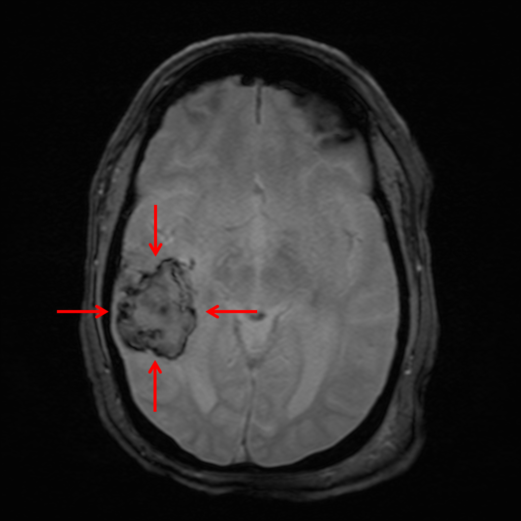
- Heterogeneous T1/T2 hyperintense lesion in the posterior right temporal lobe measuring 4.5 x 3.5 x 2 cm with associated susceptibility artifact and mild surrounding vasogenic edema with mild local mass effect resulting in partial effacement of the temporal horn of the right lateral ventricle
- Multiple small areas of restricted diffusion involving the posterior aspect of the left lentiform nucleus, left gyrus rectus, right postcentral gyrus, posterior left cingulate gyrus, right fusiform gyrus, and left cerebellar hemisphere
- Multiple foci of susceptibility artifact involving the bilateral cerebral and cerebellar hemispheres as well as the midbrain and pons
- Extensive patchy T2/FLAIR hyperintensity in the subcortical and periventricular white matter
- Age-advanced generalized cerebral atrophy
- Dilated perivascular spaces in the bilateral basal ganglia
- Polyp/mucous retention cyst in the inferior aspect of the left maxillary sinus
Diagnosis
- Hemorrhagic stroke
 Sample Report
Acute intraparenchymal hematoma in the posterior right temporal lobe measuring 4.5 x 3.5 x 2 cm (calculated volume = 16 mL) with mild surrounding vasogenic edema and resultant mild local mass effect resulting in partial effacement of the temporal horn of the right lateral ventricle. No midline shift or evidence of herniation or hydrocephalus. Given location and the presence of multiple small additional acute infarcts, this is favored to represent hemorrhagic transformation of an infarct. Hypertensive hemorrhage is thought less likely given location.
Multiple small acute infarcts involving the bilateral cerebrum and left cerebellar hemisphere without additional sites of hemorrhagic transformation.
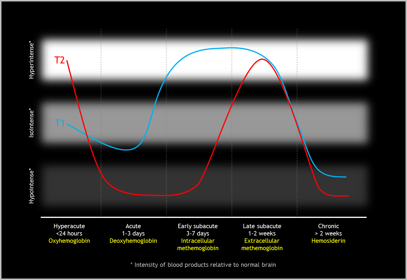
Multiple foci of susceptibility artifact involving the bilateral cerebral and cerebellar hemispheres as well as the midbrain and pons, compatible with sequela of prior microhemorrhage. While amyloid angiopathy could have this appearance, this would be atypical for patient age.
Age-advanced generalized cerebral atrophy and small vessel disease.
Discussion

📣 Feedback?
⌨️ Keyboard Shortcuts ("K")
Help | Terms | Privacy Policy | Cookie Policy
Medical Disclaimer | © 2024 CaseStacks LLC
Related Cases
References
- Bradley WG. MR appearance of hemorrhage in the brain. Radiology 1993; 189(1): 15-26.
- Larrue V, von Kummer R, Müller A, Bluhmki E. Risk Factors for Severe Hemorrhagic Transformation in Ischemic Stroke Patients Treated With Recombinant Tissue Plasminogen Activator. Stroke 2001; 32 (2): 438.
- Tomandl BF, Klotz E, Handschu R, Stemper B, Reinhardt F, Huk WJ, Eberhardt KE, Fateh-Moghadam S. Comprehensive imaging of ischemic stroke with multisection CT. Radiographics 2003; 23: 565-592.

 View shortcuts
 Zoom/Pan
 Full screen
 Window/Level
 Expand/collapse
 Scroll
 Save the case
 Close case/tab


