
Age: 62
Sex: Female
Indication: Recent stroke
Save ("V")
Case #28
Findings
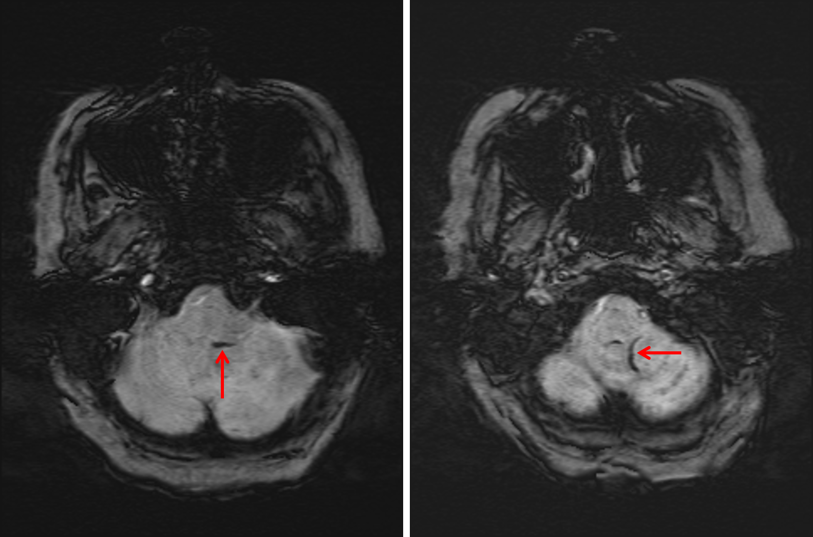
- Focus of restricted diffusion in the dorsal left aspect of the medulla
- Heterogeneous areas of faint restricted diffusion in the left cerebellar hemisphere and left occipital lobe with corresponding patchy T1 signal hyperintensity and susceptibility artifact, more confluent within the left occipital lobe
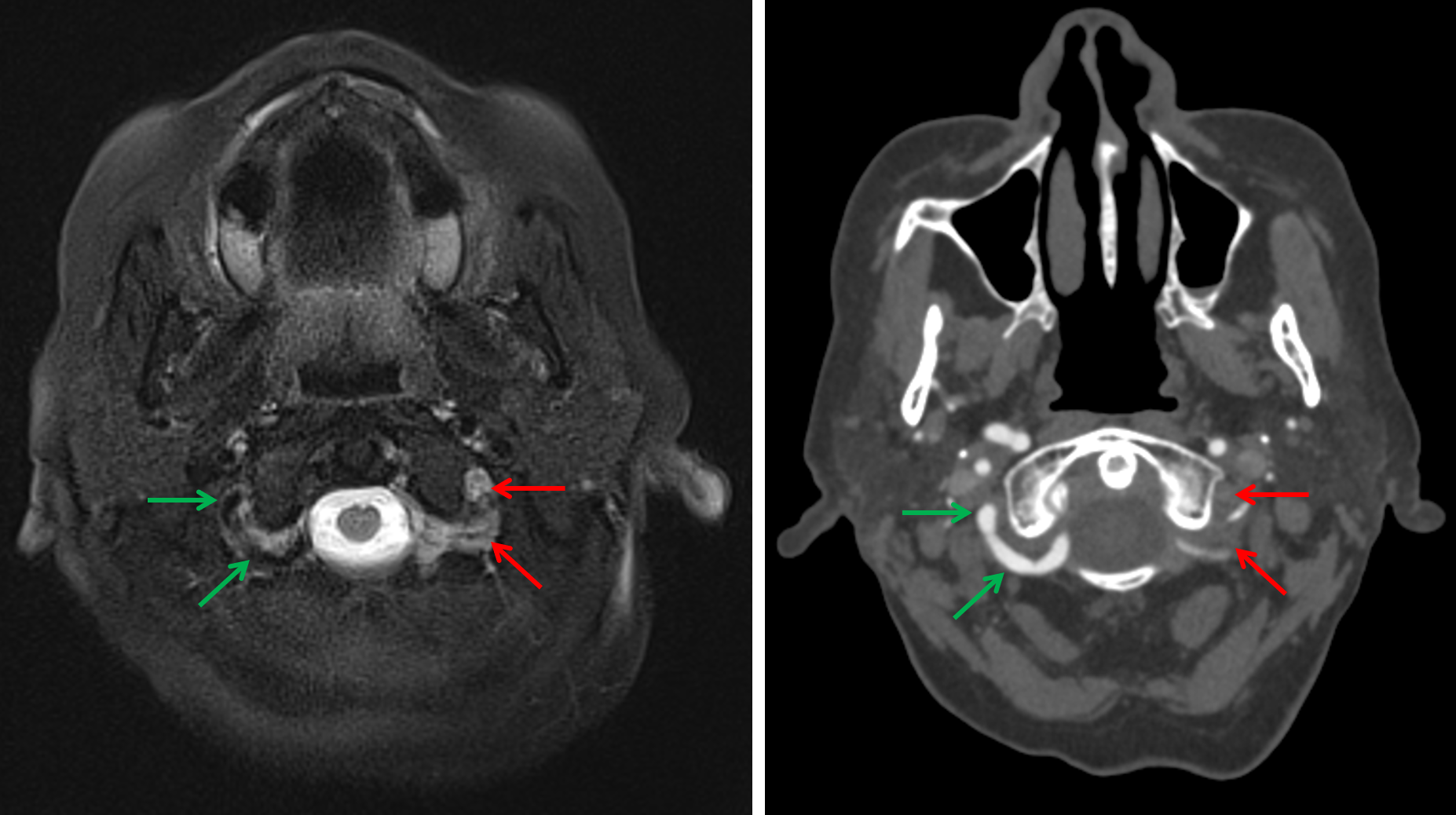
- Susceptibility artifact associated with vessels overlying the inferior aspect of the left cerebellar hemisphere
- Abnormal flow-related signal in the visualized portion of the extradural left vertebral artery
- T2/FLAIR hyperintensity in the left cerebellar hemisphere extending into the left superior and middle cerebellar peduncles with mild swelling and partial compression of the left lateral margin of the fourth ventricle
- Multiple remote lacunar infarcts involving the bilateral basal ganglia, right thalamus, left internal capsule, right aspect of the genu of the corpus callosum, and right cerebellar hemisphere
- Patchy T2/FLAIR hyperintensities in the subcortical and periventricular white matter
Diagnosis
- Infarcts with petechial hemorrhage
 Sample Report
Small acute/early subacute infarct in the left dorsal aspect of the medulla.
Subacute infarcts in the left cerebellar hemisphere and left occipital lobe with corresponding petechial hemorrhage as well as small hematoma formation in the left occipital lobe. Associated edema and mild mass effect, moreso in the left cerebellar hemisphere with mild mass effect on the fourth ventricle. No evidence of herniation or hydrocephalus.
Abnormal signal in the extradural left vertebral artery concerning for high grade stenosis or occlusion. Susceptibility artifact associated with vessels overlying the inferior aspect of the left cerebellar hemisphere is concerning for thrombus within PICA branches versus elevated deoxyhemoglobin content within cortical veins. Recommend correlation with CTA or catheter angiography.
Background of multiple remote lacunar infarcts and chronic small vessel disease.
Discussion

📣 Feedback?
⌨️ Keyboard Shortcuts ("K")
Help | Terms | Privacy Policy | Cookie Policy
Medical Disclaimer | © 2024 CaseStacks LLC
Related Cases
References
- Bradley WG. MR appearance of hemorrhage in the brain. Radiology 1993; 189(1): 15-26.
- Larrue V, von Kummer R, Müller A, Bluhmki E. Risk Factors for Severe Hemorrhagic Transformation in Ischemic Stroke Patients Treated With Recombinant Tissue Plasminogen Activator. Stroke 2001; 32 (2): 438.
- Tomandl BF, Klotz E, Handschu R, Stemper B, Reinhardt F, Huk WJ, Eberhardt KE, Fateh-Moghadam S. Comprehensive imaging of ischemic stroke with multisection CT. Radiographics 2003; 23: 565-592.

 View shortcuts
 Zoom/Pan
 Full screen
 Window/Level
 Expand/collapse
 Scroll
 Save the case
 Close case/tab


