Become a Pro Member for full access to this course and more.

View Plans   Log In
Expand ("E")   Collapse

Age: 31

Sex: Male

Indication: Headache

Save ("V")

Case #3


Findings

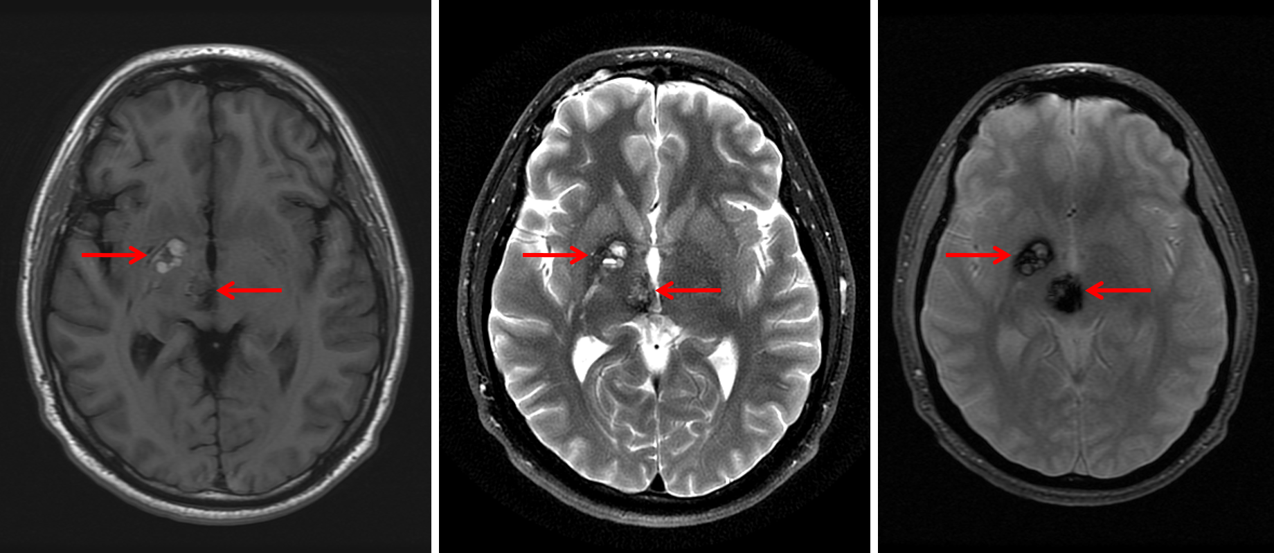
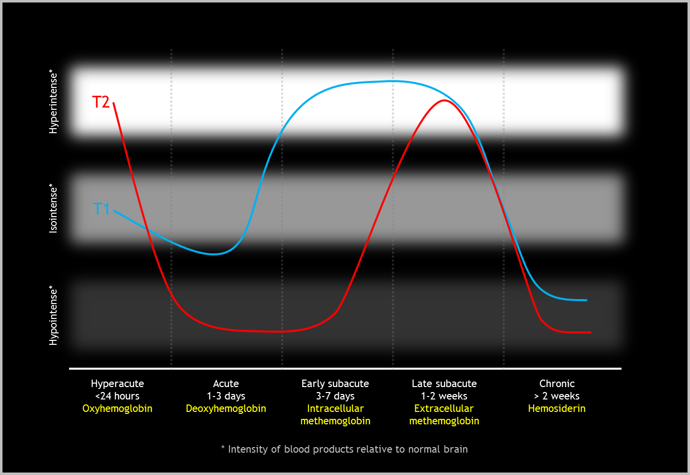
  • 2 x 1 cm lesion centered in the right globus pallidus and 1.5 x 1 cm lesion in the right midbrain, both of which have internal T1 and T2 signal hyperintensity, peripheral T2 signal hypointensity, and blooming signal hypointensity on susceptibility weighted imaging, indicating a peripheral hemosiderin ring
  • Additional small focus of susceptibility artifact in the dorsolateral right thalamus
  • The midbrain lesion exerts mild mass effect on the cerebral aqueduct without evidence of hydrocephalus
  • Developmental venous anomaly in the right thalamus, posterior limb of the right internal capsule, and posterior right lentiform nucleus draining into a right lateral tentorial sinus
  • No significant surrounding T2/FLAIR hyperintensity
  • No evidence of acute infarct
  • Few scattered T2/FLAIR hyperintensities in the cerebral white matter
  • Multiple discrete T2 signal hyperintensities and associated patchy enhancement in the superior right orbit


Diagnosis

  • Cerebral cavernous malformations (cavernomas)

Sample Report

Cerebral cavernous malformations (cavernomas) centered in the right globus pallidus and right midbrain, both of which have internal T1 and T2 signal hyperintensity suggesting subacute hemorrhage. No significant surrounding edema. The midbrain lesion exerts mild mass effect on the cerebral aqueduct without evidence of hydrocephalus. Associated developmental venous anomaly draining into a right lateral tentorial sinus.

Additional small focus of susceptibility artifact in the dorsolateral right thalamus, which could represent an additional tiny cavernoma versus sequela of prior microhemorrhage.

No evidence of acute infarct.

Few scattered T2/FLAIR hyperintensities in the cerebral white matter, which are nonspecific and may represent sequela of chronic small vessel disease or sequela of prior trauma/inflammation/vasculopathy.

Multiple discrete T2 signal hyperintensities and associated patchy enhancement in the superior right orbit, which may represent a venolymphatic malformation. Recommend dedicated orbit MRI with and without contrast.


Discussion

Become a Pro Member to unlock the discussion.

View Plans   Log In


Images

Cerebral cavenous malformations (cavernomas) centered in the right globus pallidus and right midbrain (red arrows) with associated T1 and T2 internal hyperintensity (left and middle images) and blooming on susceptibility-weighted imaging (right image). Evolution of parenchymal hematoma T1 and T2 signal over time.


📣 Feedback?

⌨️  Keyboard Shortcuts ("K")

Call Preparation

Prepare for call efficiently with interactive cases, sample reports, and annotated images.

1,400+ cases  91.25 CME

Call Simulator

   Neuro CT

250 cases

  Neuro MRI

146 cases

   Peds Radiographs

372 cases

   MSK Radiographs

281 cases

   Chest CT

84 cases

   Chest Radiographs

97 cases

   KUB

43 cases

   Body CT

140 cases

   Ultrasound

75 cases

Call Simulator

• Shuffle cases from our courses to simulate the mix of a call shift.
• Submit your own report before reviewing the case write-up.

Neuro Fellowship

Reviews of neuro topics with clinical pearls, differentials, and in-depth discussions.

131 cases  13.1 CME

   Brain Tumors

105 cases

   Neurodegenerative

13 cases

   Congenital Hearing Loss

13 cases

Call Preparation

   Neuro CT

250 cases

   Neuro MRI

146 cases

   Peds Radiographs

372 cases

   MSK Radiographs

281 cases

   Ultrasound

75 cases

   Chest CT

84 cases

   Chest Radiographs

97 cases

   Body CT

127 cases

   KUB

43 cases

Neuro Fellowship

   Brain Tumors

105 cases

   Neurodegenerative

13 cases

Call Simulator

• Shuffle cases from our courses.
• Submit your own report.

CME

• Earn up to 91 CME.
• Claim your CME to receive a certificate.

Call Simulator

• Shuffle cases from our courses to simulate the mix of a call shift.
• Submit your own report before reviewing the case write-up.

CME

• Earn up to 91 CME by completing cases in our radiology courses.
• Claim your CME to receive a certificate.

On Call

Quick references created specifically to help while on call.

  Peds Normals by Age

Reference database of normal imaging from birth to age 16

   Incidental Findings

Summary of consensus guidelines for managing incidental CT findings

   Media Index

Index of select illustrations & videos from our courses

  Neuro CT Mimics

Visual reference for common mimics of pathology on CT

On Call

  Peds Normals by Age

Reference database of normal imaging from birth to age 16

   Incidental Findings

Summary of consensus guidelines for managing incidental CT findings

   Media Index

Index of select illustrations & videos from our courses

  Neuro CT Mimics

Visual reference for common mimics of pathology on CT

Anatomy

Labelled radiographs and CT/MRI series teaching anatomy with a level of detail appropriate for medical students and junior residents.

   Pelvis

Pelvic MRI anatomy

   Chest

Chest radiograph & CT anatomy

   Body

Abdominal CT anatomy

   Cardiac

Cardiac CT anatomy

   Brain

Brain & calvarial anatomy on CT/MRI

   Cranial Nerves

Cranial nerves on MRI

   Shoulder

Shoulder MRI anatomy

   Knee

Knee MRI anatomy

   Temporal Bone

Resident/fellow-level anatomy

Anatomy

   Pelvis

Pelvic MRI anatomy

   Chest

Chest radiograph & CT anatomy

   Body

Abdominal CT anatomy

   Cardiac

Cardiac CT anatomy

   Brain

Brain & calvarial anatomy on CT/MRI

   Cranial Nerves

Cranial nerves on MRI

   Shoulder

Shoulder MRI anatomy

   Knee

Knee MRI anatomy

   Temporal Bone

Resident/fellow-level anatomy

Neuro Courses

Includes our call preparation neuro courses (Neuro CT and Neuro MRI) and our neuro fellowship courses.

   Neuro CT

250 cases

  Neuro MRI

146 cases

   Brain Tumors

105 cases

   Neurodegenerative

13 cases

   Congenital Hearing Loss

Coming soon

Neuro Courses

   Neuro CT

250 cases

   Neuro MRI

146 cases

   Brain Tumors

105 cases

   Neurodegenerative

13 cases

  Congenital Hearing Loss

Coming soon

CME

• Claim your CME to receive a certificate.

Call Simulator

• Shuffle cases from our courses to simulate the mix of a call shift.
• Submit your own report before reviewing the case write-up.

CME

• Earn up to 91 CME by completing cases in our radiology courses.
• Claim your CME to receive a certificate.

   Neuro CT

250 cases

   Neuro MRI

146 cases

   Peds Radiographs

372 cases

   MSK Radiographs

281 cases

   Ultrasound

75 cases

   Chest CT

84 cases

   Chest Radiographs

97 cases

   Body CT

140 cases

   KUB

43 cases

Call Simulator

• Shuffle cases from our courses to simulate the mix of a call shift.
• Submit your own report before reviewing the case write-up.

CME

• Earn up to 91 CME by completing cases in our radiology courses.
• Claim your CME to receive a certificate.

⌨️ Keyboard Shortcuts

View shortcuts

Zoom/Pan

  Full screen

  Window/Level

  Expand/collapse

  Scroll

  Save the case

  Close case/tab

Previous Slice (PC)

Next Slice (PC)

SHIFT Previous Slice (MAC)

 SHIFT Next Slice (MAC)

 Toggle presets

 Previous series (if multiple)

 Next series (if multiple)

  Previous case (⌘ ◄ on a mac)

  Next case (⌘ ► on a mac)

Toggle dark mode