Become a Pro Member for full access to this course and more.

View Plans   Log In
Expand ("E")   Collapse

Age: 32

Sex: Male

Indication: Trauma, right occipital condyle fracture on prior CT

Save ("V")

Case #5


Findings

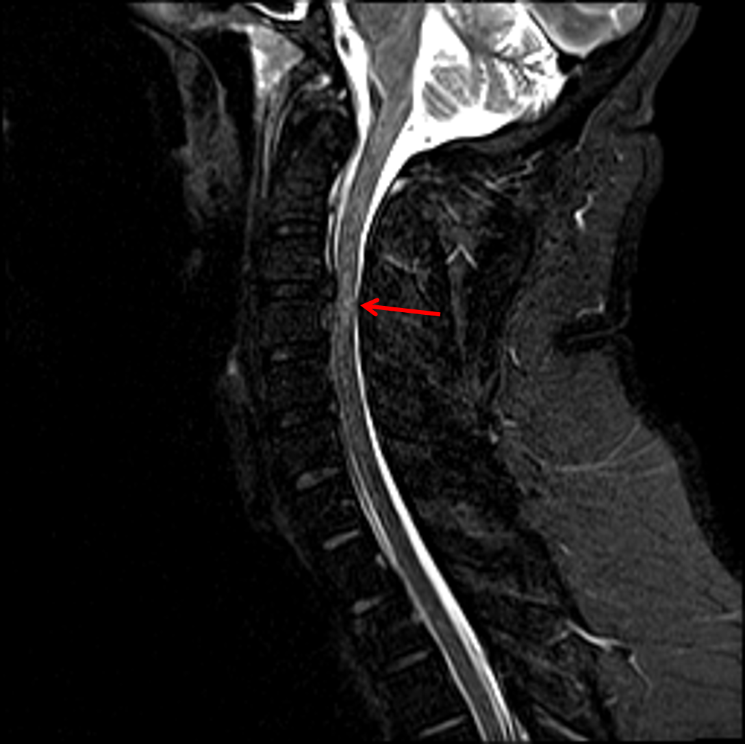
  • Large right central disc herniation at C3-C4, which contacts and deforms the right ventral cord contributing to severe right eccentric spinal canal stenosis and effacement of the right subarticular zone
  • Central cord signal abnormality at and just inferior to the C3-C4 disc herniation
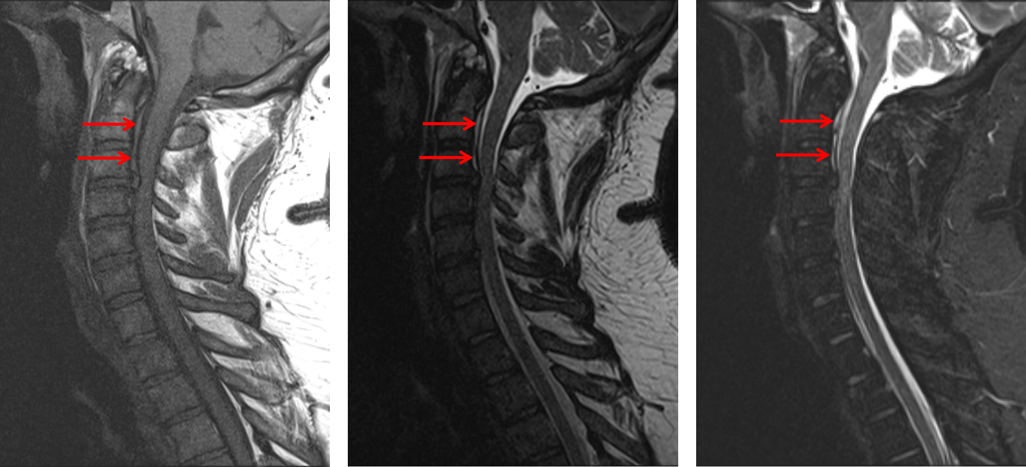
  • T1/T2 hyperintense ventral epidural collection spanning C1-C3, measuring up to 3 mm in thickness
  • No T2/STIR hyperintensity in the visualized osseous structures
  • Transverse and alar ligaments appear intact
  • Small posterior disc osteophyte complexes and uncovertebral spurring at C4-C5 and C5-C6 contribute to mild spinal canal stenosis and mild left greater than right neural foraminal stenosis
  • Mild left neural foraminal stenosis at C6-C7
  • Left dominant vertebral artery with normal flow-related signal loss in both vertebral arteries


Diagnosis

  • Traumatic disc herniation with spinal cord contusion

Sample Report

Age-indeterminate large right central disc herniation at C3-C4, which contacts and deforms the right ventral cord contributing to severe right eccentric spinal canal stenosis and effacement of the right subarticular zone. Cord signal abnormality at and just inferior to this disc herniation is concerning for contusion in the setting of trauma.

Thin ventral epidural hematoma spanning C1-C3, which measures up to 3 mm in thickness.

The reported right occipital condyle fracture is not well demonstrated on this study. Transverse and alar ligaments appear intact.


Discussion

Become a Pro Member to unlock the discussion.

View Plans   Log In


Images

Large disc herniation at C3-C4 with an associated traumatic cord contusion (red arrow). Ventral epidural collection spanning the levels of C1-C3 which appears hyperintense on both T1 (image to the left) and T2 (image in the middle) weighted sequences and does not demonstrate signal suppression on STIR (image to the right), consistent with an epidural hematoma.


📣 Feedback?

⌨️  Keyboard Shortcuts ("K")

Call Preparation

Prepare for call efficiently with interactive cases, sample reports, and annotated images.

1,400+ cases  91.25 CME

Call Simulator

   Neuro CT

250 cases

  Neuro MRI

146 cases

   Peds Radiographs

372 cases

   MSK Radiographs

281 cases

   Chest CT

84 cases

   Chest Radiographs

97 cases

   KUB

43 cases

   Body CT

140 cases

   Ultrasound

75 cases

Call Simulator

• Shuffle cases from our courses to simulate the mix of a call shift.
• Submit your own report before reviewing the case write-up.

Neuro Fellowship

Reviews of neuro topics with clinical pearls, differentials, and in-depth discussions.

131 cases  13.1 CME

   Brain Tumors

105 cases

   Neurodegenerative

13 cases

   Congenital Hearing Loss

13 cases

Call Preparation

   Neuro CT

250 cases

   Neuro MRI

146 cases

   Peds Radiographs

372 cases

   MSK Radiographs

281 cases

   Ultrasound

75 cases

   Chest CT

84 cases

   Chest Radiographs

97 cases

   Body CT

127 cases

   KUB

43 cases

Neuro Fellowship

   Brain Tumors

105 cases

   Neurodegenerative

13 cases

Call Simulator

• Shuffle cases from our courses.
• Submit your own report.

CME

• Earn up to 91 CME.
• Claim your CME to receive a certificate.

Call Simulator

• Shuffle cases from our courses to simulate the mix of a call shift.
• Submit your own report before reviewing the case write-up.

CME

• Earn up to 91 CME by completing cases in our radiology courses.
• Claim your CME to receive a certificate.

On Call

Quick references created specifically to help while on call.

  Peds Normals by Age

Reference database of normal imaging from birth to age 16

   Incidental Findings

Summary of consensus guidelines for managing incidental CT findings

   Media Index

Index of select illustrations & videos from our courses

  Neuro CT Mimics

Visual reference for common mimics of pathology on CT

On Call

  Peds Normals by Age

Reference database of normal imaging from birth to age 16

   Incidental Findings

Summary of consensus guidelines for managing incidental CT findings

   Media Index

Index of select illustrations & videos from our courses

  Neuro CT Mimics

Visual reference for common mimics of pathology on CT

Anatomy

Labelled radiographs and CT/MRI series teaching anatomy with a level of detail appropriate for medical students and junior residents.

   Pelvis

Pelvic MRI anatomy

   Chest

Chest radiograph & CT anatomy

   Body

Abdominal CT anatomy

   Cardiac

Cardiac CT anatomy

   Brain

Brain & calvarial anatomy on CT/MRI

   Cranial Nerves

Cranial nerves on MRI

   Shoulder

Shoulder MRI anatomy

   Knee

Knee MRI anatomy

   Temporal Bone

Resident/fellow-level anatomy

Anatomy

   Pelvis

Pelvic MRI anatomy

   Chest

Chest radiograph & CT anatomy

   Body

Abdominal CT anatomy

   Cardiac

Cardiac CT anatomy

   Brain

Brain & calvarial anatomy on CT/MRI

   Cranial Nerves

Cranial nerves on MRI

   Shoulder

Shoulder MRI anatomy

   Knee

Knee MRI anatomy

   Temporal Bone

Resident/fellow-level anatomy

Neuro Courses

Includes our call preparation neuro courses (Neuro CT and Neuro MRI) and our neuro fellowship courses.

   Neuro CT

250 cases

  Neuro MRI

146 cases

   Brain Tumors

105 cases

   Neurodegenerative

13 cases

   Congenital Hearing Loss

Coming soon

Neuro Courses

   Neuro CT

250 cases

   Neuro MRI

146 cases

   Brain Tumors

105 cases

   Neurodegenerative

13 cases

  Congenital Hearing Loss

Coming soon

CME

• Claim your CME to receive a certificate.

Call Simulator

• Shuffle cases from our courses to simulate the mix of a call shift.
• Submit your own report before reviewing the case write-up.

CME

• Earn up to 91 CME by completing cases in our radiology courses.
• Claim your CME to receive a certificate.

   Neuro CT

250 cases

   Neuro MRI

146 cases

   Peds Radiographs

372 cases

   MSK Radiographs

281 cases

   Ultrasound

75 cases

   Chest CT

84 cases

   Chest Radiographs

97 cases

   Body CT

140 cases

   KUB

43 cases

Call Simulator

• Shuffle cases from our courses to simulate the mix of a call shift.
• Submit your own report before reviewing the case write-up.

CME

• Earn up to 91 CME by completing cases in our radiology courses.
• Claim your CME to receive a certificate.

⌨️ Keyboard Shortcuts

View shortcuts

Zoom/Pan

  Full screen

  Window/Level

  Expand/collapse

  Scroll

  Save the case

  Close case/tab

Previous Slice (PC)

Next Slice (PC)

SHIFT Previous Slice (MAC)

 SHIFT Next Slice (MAC)

 Toggle presets

 Previous series (if multiple)

 Next series (if multiple)

  Previous case (⌘ ◄ on a mac)

  Next case (⌘ ► on a mac)

Toggle dark mode