Become a Pro Member for full access to this course and more.

View Plans   Log In
Expand ("E")   Collapse

Age: 68

Sex: Male

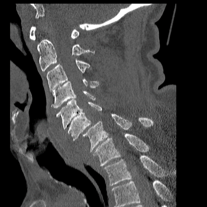
Indication: Trauma, jumped facets at C6-C7 and C4 left lamina and spinous process fracture on prior CT

Save ("V")

Case #29


Findings

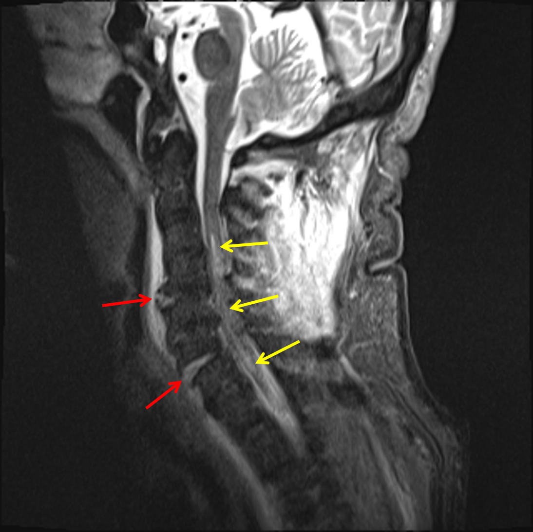
  • Mild residual anterolisthesis at C6-C7 with widening of the anterior disc space and disruption of the anterior longitudinal ligament, the posterior longitudinal ligament, and the ligamentum flavum at this level
  • Additional disruption of anterior longitudinal ligament at the level of C4-C5 as well as possible focal disruption of the posterior longitudinal ligament at C4-C5 and possible focal ligamentum flavum disruption at C3-C4
  • T2/STIR signal hyperintensity in the central aspect of the spinal cord extending from C3-C4 through C6-C7
  • T2/STIR signal hyperintensity in the bilateral C6-C7 facet joints
  • Extensive T2/STIR signal hyperintensity in the interspinous ligaments and supraspinous ligament from the skull base through C6-C7
  • Prevertebral soft tissue thickening and T2/STIR signal hyperintensity throughout the cervical spine extending into the upper thoracic spine
  • T2/STIR signal hyperintensity in the C4 left lamina and spinous process
  • Multilevel posterior disc osteophyte complexes, facet hypertrophy, uncovertebral spurring, and ligamentum flavum thickening contribute to advanced spinal canal stenosis from C4-C5 through C6-C7 and varying degrees of multilevel neural foraminal stenosis, severe bilaterally from C4-C5 through C6-C7


Diagnosis

  • Cord contusion, ligamentous injury

Sample Report

Mild residual anterolisthesis at C6-C7 with widening of the anterior disc space, evidence of bilateral facet capsular injury, and disruption of the anterior longitudinal ligament, the posterior longitudinal ligament, and the ligamentum flavum at this level. Additional disruption of anterior longitudinal ligament at the level of C4-C5 as well as possible focal disruption of the posterior longitudinal ligament at C4-C5 and possible focal ligamentum flavum disruption at C3-C4.

T2/STIR signal hyperintensity in the central aspect of the spinal cord extending from C3-C4 through C6-C7 concerning for cord contusion.

Extensive ligamentous injury involving the interspinous ligaments and supraspinous ligament from the skull base through C6-C7.

T2/STIR signal hyperintensity in the C4 left lamina and spinous process, corresponding with the known fractures.

Prevertebral soft tissue edema throughout the cervical spine extending into the upper thoracic spine.

Background of advanced degenerative changes with multilevel posterior disc osteophyte complexes, facet hypertrophy, uncovertebral spurring, and ligamentum flavum thickening contribute to advanced spinal canal stenosis from C4-C5 through C6-C7 and varying degrees of multilevel neural foraminal stenosis, severe bilaterally from C4-C5 through C6-C7.


Discussion

Become a Pro Member to unlock the discussion.

View Plans   Log In


Images

Extensive ligamentous injury in the cervical spine including focal disruptions of the anterior longitudinal ligament at two levels (red arrows). Long segment spinal cord contusion (yellow arrows). Sagittal reformatted image from the preceding CT in this patient.


📣 Feedback?

⌨️  Keyboard Shortcuts ("K")

Call Preparation

Prepare for call efficiently with interactive cases, sample reports, and annotated images.

1,400+ cases  91.25 CME

Call Simulator

   Neuro CT

250 cases

  Neuro MRI

146 cases

   Peds Radiographs

372 cases

   MSK Radiographs

281 cases

   Chest CT

84 cases

   Chest Radiographs

97 cases

   KUB

43 cases

   Body CT

140 cases

   Ultrasound

75 cases

Call Simulator

• Shuffle cases from our courses to simulate the mix of a call shift.
• Submit your own report before reviewing the case write-up.

Neuro Fellowship

Reviews of neuro topics with clinical pearls, differentials, and in-depth discussions.

131 cases  13.1 CME

   Brain Tumors

105 cases

   Neurodegenerative

13 cases

   Congenital Hearing Loss

13 cases

Call Preparation

   Neuro CT

250 cases

   Neuro MRI

146 cases

   Peds Radiographs

372 cases

   MSK Radiographs

281 cases

   Ultrasound

75 cases

   Chest CT

84 cases

   Chest Radiographs

97 cases

   Body CT

127 cases

   KUB

43 cases

Neuro Fellowship

   Brain Tumors

105 cases

   Neurodegenerative

13 cases

Call Simulator

• Shuffle cases from our courses.
• Submit your own report.

CME

• Earn up to 91 CME.
• Claim your CME to receive a certificate.

Call Simulator

• Shuffle cases from our courses to simulate the mix of a call shift.
• Submit your own report before reviewing the case write-up.

CME

• Earn up to 91 CME by completing cases in our radiology courses.
• Claim your CME to receive a certificate.

On Call

Quick references created specifically to help while on call.

  Peds Normals by Age

Reference database of normal imaging from birth to age 16

   Incidental Findings

Summary of consensus guidelines for managing incidental CT findings

   Media Index

Index of select illustrations & videos from our courses

  Neuro CT Mimics

Visual reference for common mimics of pathology on CT

On Call

  Peds Normals by Age

Reference database of normal imaging from birth to age 16

   Incidental Findings

Summary of consensus guidelines for managing incidental CT findings

   Media Index

Index of select illustrations & videos from our courses

  Neuro CT Mimics

Visual reference for common mimics of pathology on CT

Anatomy

Labelled radiographs and CT/MRI series teaching anatomy with a level of detail appropriate for medical students and junior residents.

   Pelvis

Pelvic MRI anatomy

   Chest

Chest radiograph & CT anatomy

   Body

Abdominal CT anatomy

   Cardiac

Cardiac CT anatomy

   Brain

Brain & calvarial anatomy on CT/MRI

   Cranial Nerves

Cranial nerves on MRI

   Shoulder

Shoulder MRI anatomy

   Knee

Knee MRI anatomy

   Temporal Bone

Resident/fellow-level anatomy

Anatomy

   Pelvis

Pelvic MRI anatomy

   Chest

Chest radiograph & CT anatomy

   Body

Abdominal CT anatomy

   Cardiac

Cardiac CT anatomy

   Brain

Brain & calvarial anatomy on CT/MRI

   Cranial Nerves

Cranial nerves on MRI

   Shoulder

Shoulder MRI anatomy

   Knee

Knee MRI anatomy

   Temporal Bone

Resident/fellow-level anatomy

Neuro Courses

Includes our call preparation neuro courses (Neuro CT and Neuro MRI) and our neuro fellowship courses.

   Neuro CT

250 cases

  Neuro MRI

146 cases

   Brain Tumors

105 cases

   Neurodegenerative

13 cases

   Congenital Hearing Loss

Coming soon

Neuro Courses

   Neuro CT

250 cases

   Neuro MRI

146 cases

   Brain Tumors

105 cases

   Neurodegenerative

13 cases

  Congenital Hearing Loss

Coming soon

CME

• Claim your CME to receive a certificate.

Call Simulator

• Shuffle cases from our courses to simulate the mix of a call shift.
• Submit your own report before reviewing the case write-up.

CME

• Earn up to 91 CME by completing cases in our radiology courses.
• Claim your CME to receive a certificate.

   Neuro CT

250 cases

   Neuro MRI

146 cases

   Peds Radiographs

372 cases

   MSK Radiographs

281 cases

   Ultrasound

75 cases

   Chest CT

84 cases

   Chest Radiographs

97 cases

   Body CT

140 cases

   KUB

43 cases

Call Simulator

• Shuffle cases from our courses to simulate the mix of a call shift.
• Submit your own report before reviewing the case write-up.

CME

• Earn up to 91 CME by completing cases in our radiology courses.
• Claim your CME to receive a certificate.

⌨️ Keyboard Shortcuts

View shortcuts

Zoom/Pan

  Full screen

  Window/Level

  Expand/collapse

  Scroll

  Save the case

  Close case/tab

Previous Slice (PC)

Next Slice (PC)

SHIFT Previous Slice (MAC)

 SHIFT Next Slice (MAC)

 Toggle presets

 Previous series (if multiple)

 Next series (if multiple)

  Previous case (⌘ ◄ on a mac)

  Next case (⌘ ► on a mac)

Toggle dark mode