Become a Pro Member for full access to this course and more.

View Plans   Log In
Expand ("E")   Collapse

Age: 21

Sex: Male

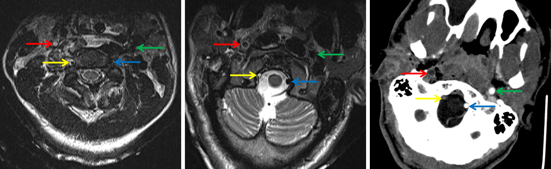
Indication: Trauma, CT showed fractures of the left occipital condyle, right C1 and C2 lateral masses, and right C3 transverse process

Save ("V")

Case #9


Findings

  • Known fractures are not well demonstrated by MRI
  • Extensive prevertebral soft tissue thickening and T2/STIR signal hyperintensity extending from the craniocervical junction to the level of C5
  • T2/STIR signal hyperintensity in the C4-C5, C5-C6, and C6-C7 interspinous ligaments with overlying T2/STIR signal hyperintensity in the posterior paraspinal soft tissues at these levels
  • Mild STIR signal hyperintensity in the supraodontoid space and right atlantooccipital joint
  • Abnormal flow-related signal in the right internal carotid artery from the level of C3 through the skull base with circumferential mural T2 signal hyperintensity in the vessel most pronounced at the level of C1
  • Abnormal flow-related signal in the right vertebral artery from the level of C1 through the distal V4 segment with areas of circumferential mural T2 signal hyperintensity more proximally in the vessel, most pronounced at the level of C3
  • T2/STIR signal hyperintensity in the right periauricular soft tissues
  • T2/STIR signal hyperintensity in the right mastoid temporal bone


Diagnosis

  • Traumatic arterial injury

Sample Report

Known fractures are not well demonstrated by MRI. No additional areas of marrow edema to suggest an additional fracture occult on CT.

Extensive prevertebral edema extending from the craniocervical junction to the level of C5 with evidence of interspinous ligament injury at C4-C5, C5-C6, and C6-C7. Mild edema in the supraodontoid space without disruption of the tectorial membrane or cruciate ligaments. Possible capsular injury involving the right atlantooccipital joint.

Abnormal flow-related signal in the right internal carotid artery from the level of C3 through the skull base and in the right vertebral artery from the level of C1 through the distal V4 segment concerning for traumatic dissection with slow flow versus occlusion. Circumferential mural T2 signal hyperintensity in both vessels raises concern for mural hematoma. Recommend CTA and/or catheter angiography for further assessment.


Discussion

Become a Pro Member to unlock the discussion.

View Plans   Log In


Images

Axial T2-weighted MR images at the level of C2 (left) and the skull base (center) and CTA axial image at the skull base (right) show abnormal flow-related signal loss and abnormal contrast filling in the right internal carotid (red arrows) and vertebral (yellow arrows) arteries compared to the normal left internal carotid (green arrows) and vertebral (blue arrows) arteries.


📣 Feedback?

⌨️  Keyboard Shortcuts ("K")

Call Preparation

Prepare for call efficiently with interactive cases, sample reports, and annotated images.

1,400+ cases  91.25 CME

Call Simulator

   Neuro CT

250 cases

  Neuro MRI

146 cases

   Peds Radiographs

372 cases

   MSK Radiographs

281 cases

   Chest CT

84 cases

   Chest Radiographs

97 cases

   KUB

43 cases

   Body CT

140 cases

   Ultrasound

75 cases

Call Simulator

• Shuffle cases from our courses to simulate the mix of a call shift.
• Submit your own report before reviewing the case write-up.

Neuro Fellowship

Reviews of neuro topics with clinical pearls, differentials, and in-depth discussions.

131 cases  13.1 CME

   Brain Tumors

105 cases

   Neurodegenerative

13 cases

   Congenital Hearing Loss

13 cases

Call Preparation

   Neuro CT

250 cases

   Neuro MRI

146 cases

   Peds Radiographs

372 cases

   MSK Radiographs

281 cases

   Ultrasound

75 cases

   Chest CT

84 cases

   Chest Radiographs

97 cases

   Body CT

127 cases

   KUB

43 cases

Neuro Fellowship

   Brain Tumors

105 cases

   Neurodegenerative

13 cases

Call Simulator

• Shuffle cases from our courses.
• Submit your own report.

CME

• Earn up to 91 CME.
• Claim your CME to receive a certificate.

Call Simulator

• Shuffle cases from our courses to simulate the mix of a call shift.
• Submit your own report before reviewing the case write-up.

CME

• Earn up to 91 CME by completing cases in our radiology courses.
• Claim your CME to receive a certificate.

On Call

Quick references created specifically to help while on call.

  Peds Normals by Age

Reference database of normal imaging from birth to age 16

   Incidental Findings

Summary of consensus guidelines for managing incidental CT findings

   Media Index

Index of select illustrations & videos from our courses

  Neuro CT Mimics

Visual reference for common mimics of pathology on CT

On Call

  Peds Normals by Age

Reference database of normal imaging from birth to age 16

   Incidental Findings

Summary of consensus guidelines for managing incidental CT findings

   Media Index

Index of select illustrations & videos from our courses

  Neuro CT Mimics

Visual reference for common mimics of pathology on CT

Anatomy

Labelled radiographs and CT/MRI series teaching anatomy with a level of detail appropriate for medical students and junior residents.

   Pelvis

Pelvic MRI anatomy

   Chest

Chest radiograph & CT anatomy

   Body

Abdominal CT anatomy

   Cardiac

Cardiac CT anatomy

   Brain

Brain & calvarial anatomy on CT/MRI

   Cranial Nerves

Cranial nerves on MRI

   Shoulder

Shoulder MRI anatomy

   Knee

Knee MRI anatomy

   Temporal Bone

Resident/fellow-level anatomy

Anatomy

   Pelvis

Pelvic MRI anatomy

   Chest

Chest radiograph & CT anatomy

   Body

Abdominal CT anatomy

   Cardiac

Cardiac CT anatomy

   Brain

Brain & calvarial anatomy on CT/MRI

   Cranial Nerves

Cranial nerves on MRI

   Shoulder

Shoulder MRI anatomy

   Knee

Knee MRI anatomy

   Temporal Bone

Resident/fellow-level anatomy

Neuro Courses

Includes our call preparation neuro courses (Neuro CT and Neuro MRI) and our neuro fellowship courses.

   Neuro CT

250 cases

  Neuro MRI

146 cases

   Brain Tumors

105 cases

   Neurodegenerative

13 cases

   Congenital Hearing Loss

Coming soon

Neuro Courses

   Neuro CT

250 cases

   Neuro MRI

146 cases

   Brain Tumors

105 cases

   Neurodegenerative

13 cases

  Congenital Hearing Loss

Coming soon

CME

• Claim your CME to receive a certificate.

Call Simulator

• Shuffle cases from our courses to simulate the mix of a call shift.
• Submit your own report before reviewing the case write-up.

CME

• Earn up to 91 CME by completing cases in our radiology courses.
• Claim your CME to receive a certificate.

   Neuro CT

250 cases

   Neuro MRI

146 cases

   Peds Radiographs

372 cases

   MSK Radiographs

281 cases

   Ultrasound

75 cases

   Chest CT

84 cases

   Chest Radiographs

97 cases

   Body CT

140 cases

   KUB

43 cases

Call Simulator

• Shuffle cases from our courses to simulate the mix of a call shift.
• Submit your own report before reviewing the case write-up.

CME

• Earn up to 91 CME by completing cases in our radiology courses.
• Claim your CME to receive a certificate.

⌨️ Keyboard Shortcuts

View shortcuts

Zoom/Pan

  Full screen

  Window/Level

  Expand/collapse

  Scroll

  Save the case

  Close case/tab

Previous Slice (PC)

Next Slice (PC)

SHIFT Previous Slice (MAC)

 SHIFT Next Slice (MAC)

 Toggle presets

 Previous series (if multiple)

 Next series (if multiple)

  Previous case (⌘ ◄ on a mac)

  Next case (⌘ ► on a mac)

Toggle dark mode