Become a Pro Member for full access to this course and more.

View Plans   Log In
Expand ("E")   Collapse

Age: 78

Sex: Male

Indication: Tarry stools, syncope

Save ("V")

Case #21


Findings

  • Lower chest
    • Advanced destructive emphysema
    • Heavy coronary artery calcification
  • Abdomen/Pelvis
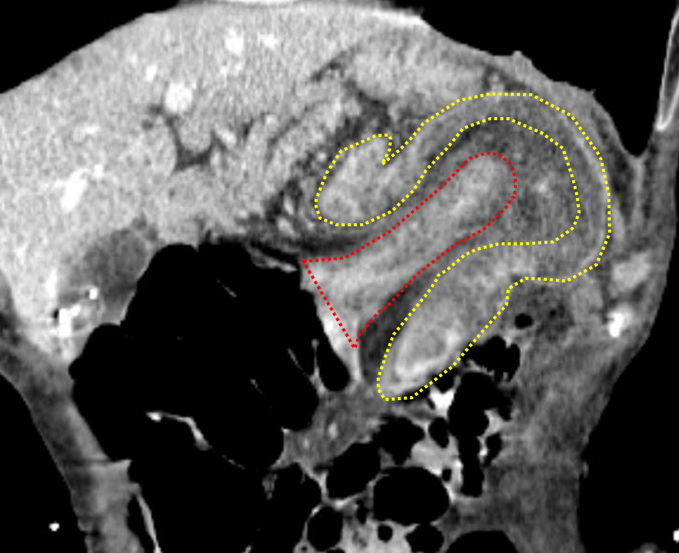
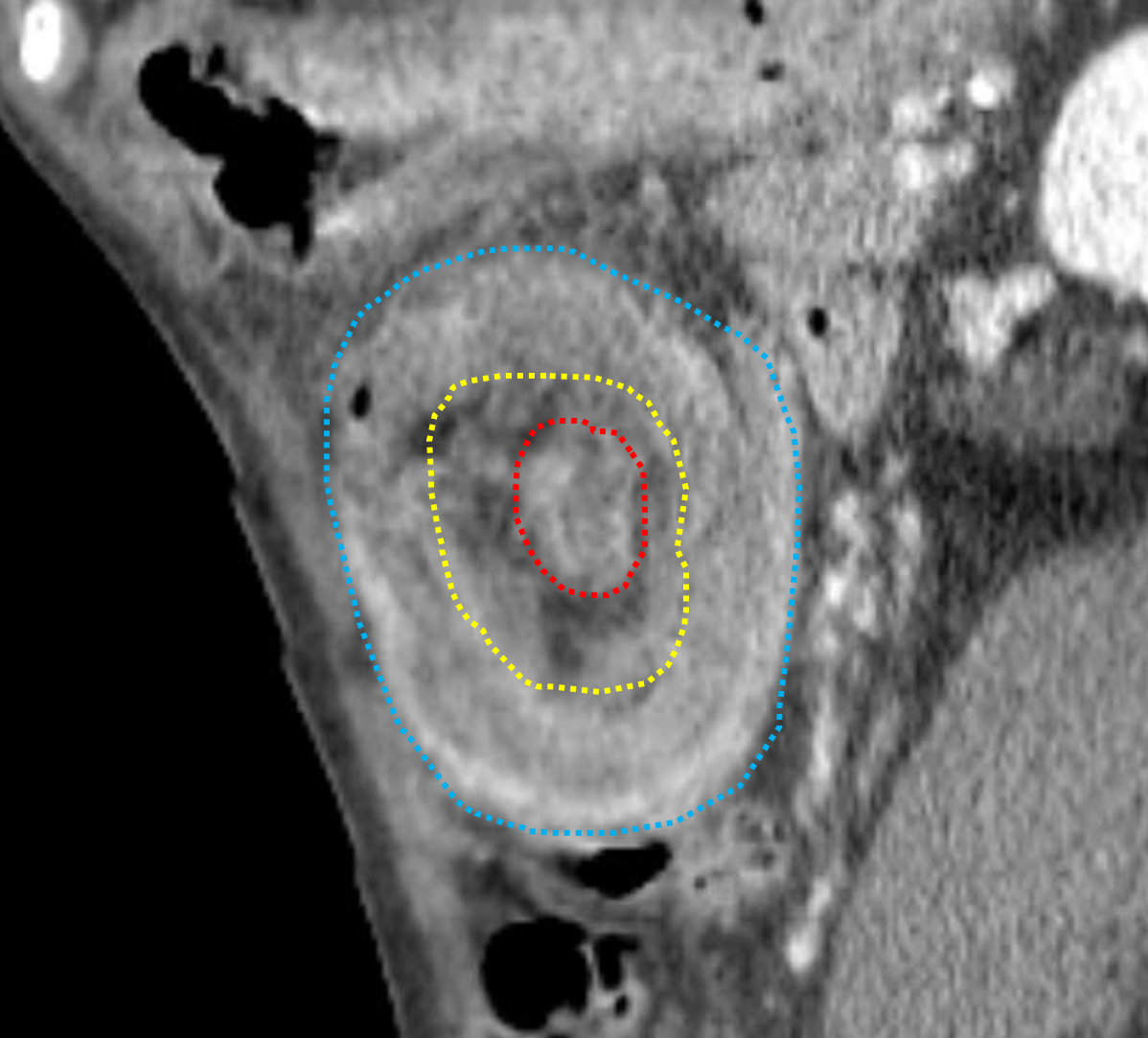
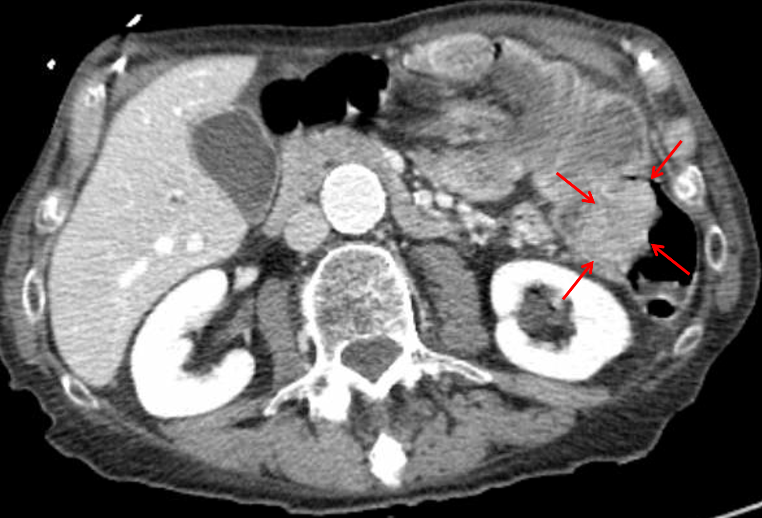
    • Colo-colonic intussusception at the splenic flexure measuring 10 cm in length with a 3.5 x 2.5 cm mass at the distal margin of the intussusceptum
    • Edematous appearance of the intussusceptum
    • Colonic diverticulosis
    • Hepatomegaly
    • Multiple circumscribed fluid attenuation hepatic lesions, likely cysts
    • Focal fatty infiltration in the liver adjacent to the falciform ligament
    • 7 mm hypodense lesion with associated calcification in the pancreatic head
    • Focal mural thickening at the gallbladder fundus compatible with adenomyomatosis
    • Mild fullness of both renal collecting systems
    • Bilateral subcentimeter renal hypodensities which are too small to characterize
    • Small volume ascites around the liver extending into the right paracolic gutter and into the anatomic pelvis
    • Distended urinary bladder
    • Prostatomegaly with nonspecific prostatic calcification
    • Atherosclerotic calcification of the abdominal aorta and branch vessels with fusiform ectasia of the infrarenal abdominal aorta measuring 2.5 cm in AP diameter
  • MSK
    • No acute osseous findings
    • Polyarticular degenerative changes
    • Body wall edema


Diagnosis

  • Malignant colo-colonic intussusception

Sample Report

Colo-colonic intussusception at the splenic flexure measuring 10 cm in length with edema and possible early ischemic changes of the intussusceptum. Mass at the distal margin of the intussusceptum is concerning for a neoplastic lead point. Recommend surgical evaluation.

Small volume free intraperitoneal fluid, likely reactive to the intussusception.

Findings of chronic bladder outlet obstruction related to prostatomegaly with mild fullness of both renal collecting systems.

7 mm partially calcified lesion in the pancreatic head. Recommend correlation with prior imaging, if available, to document stability. If no prior imaging is available, recommend followup pancreatic protocol CT or MRI in 2 years.


Discussion

Become a Pro Member to unlock the discussion.

View Plans   Log In


Images

Intussusception: coronal reformat shows the intussusceptum (outlined in red) telescoping into the intussuscepiens (outlined in yellow). Between the two is the mesentery associated with the intussusceptum. Intussusception: sagittal reformat shows the classic targetoid appearance with the intussusceptum in the center (outlined in red) surrounded by its mesentery (circled in yellow) surrounded by the intussuscepiens (circled in blue). Mass at the distal margin of the intussusceptum (red arrows) concerning for neoplastic lead point.


📣 Feedback?

⌨️  Keyboard Shortcuts ("K")

Call Preparation

Prepare for call efficiently with interactive cases, sample reports, and annotated images.

1,400+ cases  91.25 CME

Call Simulator

   Neuro CT

250 cases

  Neuro MRI

146 cases

   Peds Radiographs

372 cases

   MSK Radiographs

281 cases

   Chest CT

84 cases

   Chest Radiographs

97 cases

   KUB

43 cases

   Body CT

140 cases

   Ultrasound

75 cases

Call Simulator

• Shuffle cases from our courses to simulate the mix of a call shift.
• Submit your own report before reviewing the case write-up.

Neuro Fellowship

Reviews of neuro topics with clinical pearls, differentials, and in-depth discussions.

131 cases  13.1 CME

   Brain Tumors

105 cases

   Neurodegenerative

13 cases

   Congenital Hearing Loss

13 cases

Call Preparation

   Neuro CT

250 cases

   Neuro MRI

146 cases

   Peds Radiographs

372 cases

   MSK Radiographs

281 cases

   Ultrasound

75 cases

   Chest CT

84 cases

   Chest Radiographs

97 cases

   Body CT

127 cases

   KUB

43 cases

Neuro Fellowship

   Brain Tumors

105 cases

   Neurodegenerative

13 cases

Call Simulator

• Shuffle cases from our courses.
• Submit your own report.

CME

• Earn up to 91 CME.
• Claim your CME to receive a certificate.

Call Simulator

• Shuffle cases from our courses to simulate the mix of a call shift.
• Submit your own report before reviewing the case write-up.

CME

• Earn up to 91 CME by completing cases in our radiology courses.
• Claim your CME to receive a certificate.

On Call

Quick references created specifically to help while on call.

  Peds Normals by Age

Reference database of normal imaging from birth to age 16

   Incidental Findings

Summary of consensus guidelines for managing incidental CT findings

   Media Index

Index of select illustrations & videos from our courses

  Neuro CT Mimics

Visual reference for common mimics of pathology on CT

On Call

  Peds Normals by Age

Reference database of normal imaging from birth to age 16

   Incidental Findings

Summary of consensus guidelines for managing incidental CT findings

   Media Index

Index of select illustrations & videos from our courses

  Neuro CT Mimics

Visual reference for common mimics of pathology on CT

Anatomy

Labelled radiographs and CT/MRI series teaching anatomy with a level of detail appropriate for medical students and junior residents.

   Pelvis

Pelvic MRI anatomy

   Chest

Chest radiograph & CT anatomy

   Body

Abdominal CT anatomy

   Cardiac

Cardiac CT anatomy

   Brain

Brain & calvarial anatomy on CT/MRI

   Cranial Nerves

Cranial nerves on MRI

   Shoulder

Shoulder MRI anatomy

   Knee

Knee MRI anatomy

   Temporal Bone

Resident/fellow-level anatomy

Anatomy

   Pelvis

Pelvic MRI anatomy

   Chest

Chest radiograph & CT anatomy

   Body

Abdominal CT anatomy

   Cardiac

Cardiac CT anatomy

   Brain

Brain & calvarial anatomy on CT/MRI

   Cranial Nerves

Cranial nerves on MRI

   Shoulder

Shoulder MRI anatomy

   Knee

Knee MRI anatomy

   Temporal Bone

Resident/fellow-level anatomy

Neuro Courses

Includes our call preparation neuro courses (Neuro CT and Neuro MRI) and our neuro fellowship courses.

   Neuro CT

250 cases

  Neuro MRI

146 cases

   Brain Tumors

105 cases

   Neurodegenerative

13 cases

   Congenital Hearing Loss

Coming soon

Neuro Courses

   Neuro CT

250 cases

   Neuro MRI

146 cases

   Brain Tumors

105 cases

   Neurodegenerative

13 cases

  Congenital Hearing Loss

Coming soon

CME

• Claim your CME to receive a certificate.

Call Simulator

• Shuffle cases from our courses to simulate the mix of a call shift.
• Submit your own report before reviewing the case write-up.

CME

• Earn up to 91 CME by completing cases in our radiology courses.
• Claim your CME to receive a certificate.

   Neuro CT

250 cases

   Neuro MRI

146 cases

   Peds Radiographs

372 cases

   MSK Radiographs

281 cases

   Ultrasound

75 cases

   Chest CT

84 cases

   Chest Radiographs

97 cases

   Body CT

140 cases

   KUB

43 cases

Call Simulator

• Shuffle cases from our courses to simulate the mix of a call shift.
• Submit your own report before reviewing the case write-up.

CME

• Earn up to 91 CME by completing cases in our radiology courses.
• Claim your CME to receive a certificate.

⌨️ Keyboard Shortcuts

View shortcuts

Zoom/Pan

  Full screen

  Window/Level

  Expand/collapse

  Scroll

  Save the case

  Close case/tab

Previous Slice (PC)

Next Slice (PC)

SHIFT Previous Slice (MAC)

 SHIFT Next Slice (MAC)

 Toggle presets

 Previous series (if multiple)

 Next series (if multiple)

  Previous case (⌘ ◄ on a mac)

  Next case (⌘ ► on a mac)

Toggle dark mode