Become a Pro Member for full access to this course and more.

View Plans   Log In
Expand ("E")   Collapse

Age: 77

Sex: Female

Indication: Upper abdominal pain, fever

Save ("V")

Case #83


Findings

  • Lower chest
    • Pulmonary nodule in the medial aspect of the left lower lobe measuring 2 x 1.5 cm
    • No pleural effusions
    • Small hiatal hernia
  • Abdomen/Pelvis
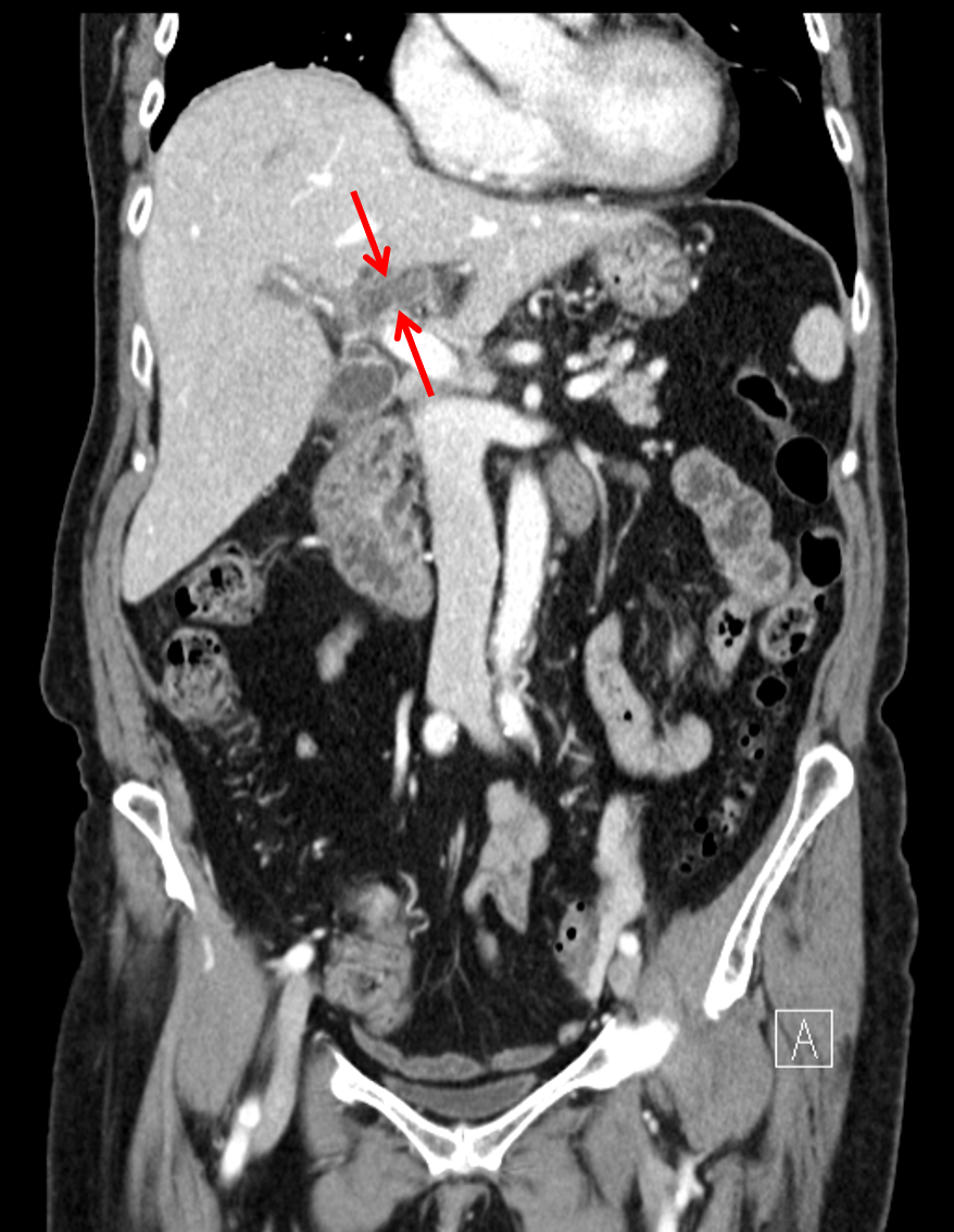
    • Occlusive thrombosis of the left portal venous system with wall thickening and hyperenhancement of the thrombosed veins
    • Thrombus also involves right portal venous branches to a lesser extent
    • The main portal vein, splenic vein, and SMV remain patent
    • Heterogeneous enhancement of the hepatic parenchyma
    • Mural thickening of the gallbladder and extrahepatic bile ducts, which are nondilated
    • Punctate calcified gallstone layering in the gallbladder fundus
    • Mildly prominent lymph node in the gastrohepatic ligament measuring 8 mm in short axis
    • Colonic diverticulosis and small periampullary duodenal diverticulum without evidence for acute diverticulitis
    • Atherosclerotic calcification of the abdominal aorta and branch vessels without aneurysm
  • MSK
    • No acute findings
    • Peripherally calcified bilateral breast implants with areas of intracapsular rupture bilaterally


Diagnosis

  • Pylephlebitis

Sample Report

Occlusive thrombosis of the left portal venous tree with associated venous wall thickening and enhancement, suspicious for thrombophlebitis (pylephlebitis). Additional areas of thrombus involving right portal venous branches. The main portal vein, splenic vein, and SMV remain patent.

Gallbladder and extrahepatic biliary duct wall thickening, which may relate to cholecystitis and cholangitis. No evidence for biliary obstruction.

Heterogeneous enhancement of the hepatic parenchyma, which may relate to altered perfusion in in the setting of portal venous thrombosis, though hepatitis could have a similar appearance. No evidence for hepatic abscess.

Left lower lobe pulmonary nodule measuring 2 cm, which is concerning for primary or metastatic malignancy. Recommend chest CT for further assessment.


Discussion

Become a Pro Member to unlock the discussion.

View Plans   Log In


Images

Left portal vein thrombosis with wall thickening and enhancement (red arrows), suspicious for pylephlebitis.


📣 Feedback?

⌨️  Keyboard Shortcuts ("K")

Call Preparation

Prepare for call efficiently with interactive cases, sample reports, and annotated images.

1,400+ cases  91.25 CME

Call Simulator

   Neuro CT

250 cases

  Neuro MRI

146 cases

   Peds Radiographs

372 cases

   MSK Radiographs

281 cases

   Chest CT

84 cases

   Chest Radiographs

97 cases

   KUB

43 cases

   Body CT

140 cases

   Ultrasound

75 cases

Call Simulator

• Shuffle cases from our courses to simulate the mix of a call shift.
• Submit your own report before reviewing the case write-up.

Neuro Fellowship

Reviews of neuro topics with clinical pearls, differentials, and in-depth discussions.

131 cases  13.1 CME

   Brain Tumors

105 cases

   Neurodegenerative

13 cases

   Congenital Hearing Loss

13 cases

Call Preparation

   Neuro CT

250 cases

   Neuro MRI

146 cases

   Peds Radiographs

372 cases

   MSK Radiographs

281 cases

   Ultrasound

75 cases

   Chest CT

84 cases

   Chest Radiographs

97 cases

   Body CT

127 cases

   KUB

43 cases

Neuro Fellowship

   Brain Tumors

105 cases

   Neurodegenerative

13 cases

Call Simulator

• Shuffle cases from our courses.
• Submit your own report.

CME

• Earn up to 91 CME.
• Claim your CME to receive a certificate.

Call Simulator

• Shuffle cases from our courses to simulate the mix of a call shift.
• Submit your own report before reviewing the case write-up.

CME

• Earn up to 91 CME by completing cases in our radiology courses.
• Claim your CME to receive a certificate.

On Call

Quick references created specifically to help while on call.

  Peds Normals by Age

Reference database of normal imaging from birth to age 16

   Incidental Findings

Summary of consensus guidelines for managing incidental CT findings

   Media Index

Index of select illustrations & videos from our courses

  Neuro CT Mimics

Visual reference for common mimics of pathology on CT

On Call

  Peds Normals by Age

Reference database of normal imaging from birth to age 16

   Incidental Findings

Summary of consensus guidelines for managing incidental CT findings

   Media Index

Index of select illustrations & videos from our courses

  Neuro CT Mimics

Visual reference for common mimics of pathology on CT

Anatomy

Labelled radiographs and CT/MRI series teaching anatomy with a level of detail appropriate for medical students and junior residents.

   Pelvis

Pelvic MRI anatomy

   Chest

Chest radiograph & CT anatomy

   Body

Abdominal CT anatomy

   Cardiac

Cardiac CT anatomy

   Brain

Brain & calvarial anatomy on CT/MRI

   Cranial Nerves

Cranial nerves on MRI

   Shoulder

Shoulder MRI anatomy

   Knee

Knee MRI anatomy

   Temporal Bone

Resident/fellow-level anatomy

Anatomy

   Pelvis

Pelvic MRI anatomy

   Chest

Chest radiograph & CT anatomy

   Body

Abdominal CT anatomy

   Cardiac

Cardiac CT anatomy

   Brain

Brain & calvarial anatomy on CT/MRI

   Cranial Nerves

Cranial nerves on MRI

   Shoulder

Shoulder MRI anatomy

   Knee

Knee MRI anatomy

   Temporal Bone

Resident/fellow-level anatomy

Neuro Courses

Includes our call preparation neuro courses (Neuro CT and Neuro MRI) and our neuro fellowship courses.

   Neuro CT

250 cases

  Neuro MRI

146 cases

   Brain Tumors

105 cases

   Neurodegenerative

13 cases

   Congenital Hearing Loss

Coming soon

Neuro Courses

   Neuro CT

250 cases

   Neuro MRI

146 cases

   Brain Tumors

105 cases

   Neurodegenerative

13 cases

  Congenital Hearing Loss

Coming soon

CME

• Claim your CME to receive a certificate.

Call Simulator

• Shuffle cases from our courses to simulate the mix of a call shift.
• Submit your own report before reviewing the case write-up.

CME

• Earn up to 91 CME by completing cases in our radiology courses.
• Claim your CME to receive a certificate.

   Neuro CT

250 cases

   Neuro MRI

146 cases

   Peds Radiographs

372 cases

   MSK Radiographs

281 cases

   Ultrasound

75 cases

   Chest CT

84 cases

   Chest Radiographs

97 cases

   Body CT

140 cases

   KUB

43 cases

Call Simulator

• Shuffle cases from our courses to simulate the mix of a call shift.
• Submit your own report before reviewing the case write-up.

CME

• Earn up to 91 CME by completing cases in our radiology courses.
• Claim your CME to receive a certificate.

⌨️ Keyboard Shortcuts

View shortcuts

Zoom/Pan

  Full screen

  Window/Level

  Expand/collapse

  Scroll

  Save the case

  Close case/tab

Previous Slice (PC)

Next Slice (PC)

SHIFT Previous Slice (MAC)

 SHIFT Next Slice (MAC)

 Toggle presets

 Previous series (if multiple)

 Next series (if multiple)

  Previous case (⌘ ◄ on a mac)

  Next case (⌘ ► on a mac)

Toggle dark mode