Expand ("E")   Collapse

Age: 59

Sex: Male

Indication: Trauma

Save ("V")

Case #1


Findings

  • Chest
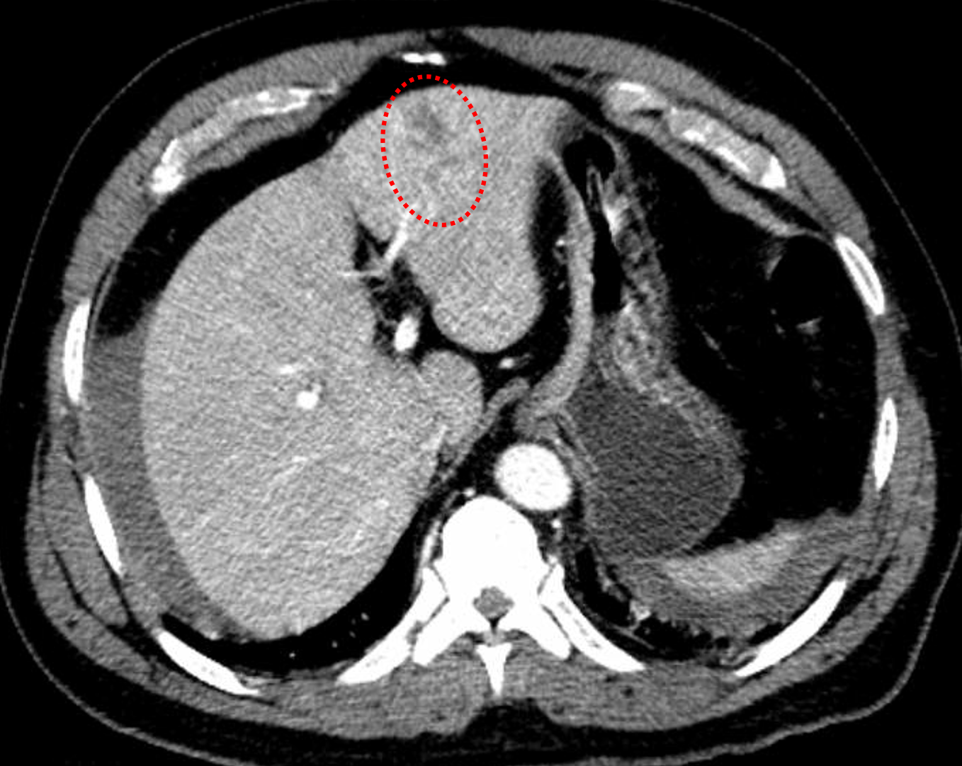
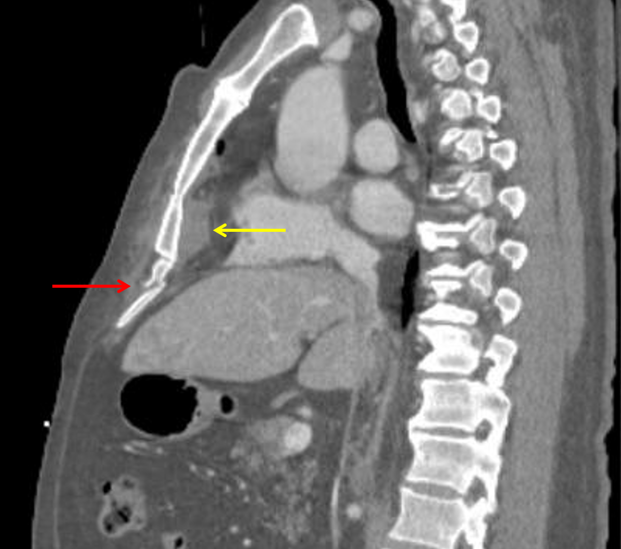
    • Small lower anterior mediastinal hematoma without active extravasation
    • Trace right pneumothorax
    • Mild contusion in the anterior aspect of the left upper lobe
    • Mild dependent bilataral lower lobe atelectasis
    • Small hiatal hernia
  • Abdomen/Pelvis
    • Hepatic laceration involving segments 2/3 measuring 4.4 cm in greatest span. No extension to the hilum or evidence of active hemorrhage
    • Moderate volume hemoperitoneum, particularly concentrated about the liver and in the right paracolic gutter
    • Stranding in the right ileocolic and left colic mesentery with areas of associated contrast density which change in size and appearance on delayed images
  • MSK
    • Acute nondisplaced fractures of the right lateral third and fourth ribs
    • Acute minimally displaced fractures through the costal cartilage of multiple right ribs
    • Remote right lateral eighth and ninth rib fractures
    • Acute minimally displaced fracture of the xiphoid process
    • Acute minimally displaced fracture of the anterior superior corner of the L4 vertebral body.
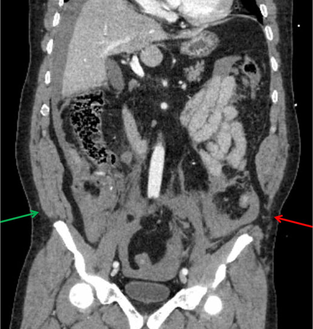
    • Traumatic rupture of all fascial layers of the left lateral abdominal wall near their attachment on the left iliac crest resulting in a lumbar hernia, which only contains fat
    • Intramuscular hematoma in the imaged portion of the left sternocleiodomastoid muscle
    • Soft tissue contusion anterior to the sternum


Diagnosis

  • Hepatic laceration
  • Mesenteric injuries with active hemorrhage
  • Mediastinal hematoma
  • Traumatic lumbar hernia

Sample Report

Traumatic hematomas in the right ileocolic and left colic mesenteries with evidence of active hemorrhage at both locations.

Grade 3 hepatic laceration in segments 2/3 with moderate volume hemoperitoneum.

Acute left lumbar hernia with herniation of abdominal fat through the defect.

Acute fractures through the right costal cartilage and xiphoid process with a small lower mediastinal hematoma. No direct evidence of acute aortic injury.

Acute nondisplaced fractures of the right lateral third and fourth ribs with a tiny right pneumothorax.

Acute minimally displaced fracture of the anterior superior corner of the L4 vertebral body.

Partially imaged intramuscular hematoma in the left sternocleidomastoid muscle.


Discussion

  • Hepatic injuries can be classified using the AAST system, as shown in the table below. While including the classification of injuries is not always important, it is important to generate reports that will be meaningful to your referring surgeons.
  • Any time you see a laceration or hematoma, make sure to compare closely between arterial and delayed phases for signs of arterial injury including hemorrhage (should get larger and less poorly defined on delayed phase) and pseudoaneurysm (should remain similar in appearance on delayed phase)
  • When you see a laceration that extends to or near the hepatic hilum, make sure to look for biliary complications which can include:
    • Hemorrhage into a bile duct causing obstruction – look for regional intrahepatic duct dilation
    • Bile leak, which will initially look like nonloculated low density fluid, but will progress to a biloma over time
  • This case also shows a good example of a traumatic lumbar hernia, as shown in an annotated image below. While rare, these indicate severe shearing forces and can unfortunately easily be missed by clinicians and radiologists in polytrauma cases


AAST Liver Injury Scale

Grade Laceration Subcapsular hematoma Parenchymal hematoma Major vascular injury
I < 1 cm in depth < 10% of the hepatic surface area None None
II 1-3 cm in depth 10-50% of the hepatic surface area < 10 cm in diameter None
III > 3 cm in depth > 50% of the hepatic surface area
Expanding
Ruptured
> 10 cm in diameter
Expanding
Ruptured
Active bleeding confined within the liver
IV Disruption of 25-75% of a lobe (or 1-3 segments of a lobe) Any Any Active bleeding extending beyond the liver
V Disruption of > 75% of a lobe (or > 3 segments of a lobe) Any Any Juxtahepatic venous injury (IVC or central hepatic veins)
VI Any Any Any Avulsion of the hepatic hilum


Images

Red outline: hepatic laceration in segment 4 measuring greater than 3 cm in depth. Xiphoid process fracture (red arrow) and subjacent mediastinal hematoma (yellow arrow). Traumatic herniation of the abdominal wall musculature from the left iliac crest (red arrow) compared to the normal appearance on the right (green arrow).


📣 Feedback?

⌨️  Keyboard Shortcuts ("K")

Call Preparation

Prepare for call efficiently with interactive cases, sample reports, and annotated images.

1,400+ cases  91.25 CME

Call Simulator

   Neuro CT

250 cases

  Neuro MRI

146 cases

   Peds Radiographs

372 cases

   MSK Radiographs

281 cases

   Chest CT

84 cases

   Chest Radiographs

97 cases

   KUB

43 cases

   Body CT

140 cases

   Ultrasound

75 cases

Call Simulator

• Shuffle cases from our courses to simulate the mix of a call shift.
• Submit your own report before reviewing the case write-up.

Neuro Fellowship

Reviews of neuro topics with clinical pearls, differentials, and in-depth discussions.

131 cases  13.1 CME

   Brain Tumors

105 cases

   Neurodegenerative

13 cases

   Congenital Hearing Loss

13 cases

Call Preparation

   Neuro CT

250 cases

   Neuro MRI

146 cases

   Peds Radiographs

372 cases

   MSK Radiographs

281 cases

   Ultrasound

75 cases

   Chest CT

84 cases

   Chest Radiographs

97 cases

   Body CT

127 cases

   KUB

43 cases

Neuro Fellowship

   Brain Tumors

105 cases

   Neurodegenerative

13 cases

Call Simulator

• Shuffle cases from our courses.
• Submit your own report.

CME

• Earn up to 91 CME.
• Claim your CME to receive a certificate.

Call Simulator

• Shuffle cases from our courses to simulate the mix of a call shift.
• Submit your own report before reviewing the case write-up.

CME

• Earn up to 91 CME by completing cases in our radiology courses.
• Claim your CME to receive a certificate.

On Call

Quick references created specifically to help while on call.

  Peds Normals by Age

Reference database of normal imaging from birth to age 16

   Incidental Findings

Summary of consensus guidelines for managing incidental CT findings

   Media Index

Index of select illustrations & videos from our courses

  Neuro CT Mimics

Visual reference for common mimics of pathology on CT

On Call

  Peds Normals by Age

Reference database of normal imaging from birth to age 16

   Incidental Findings

Summary of consensus guidelines for managing incidental CT findings

   Media Index

Index of select illustrations & videos from our courses

  Neuro CT Mimics

Visual reference for common mimics of pathology on CT

Anatomy

Labelled radiographs and CT/MRI series teaching anatomy with a level of detail appropriate for medical students and junior residents.

   Pelvis

Pelvic MRI anatomy

   Chest

Chest radiograph & CT anatomy

   Body

Abdominal CT anatomy

   Cardiac

Cardiac CT anatomy

   Brain

Brain & calvarial anatomy on CT/MRI

   Cranial Nerves

Cranial nerves on MRI

   Shoulder

Shoulder MRI anatomy

   Knee

Knee MRI anatomy

   Temporal Bone

Resident/fellow-level anatomy

Anatomy

   Pelvis

Pelvic MRI anatomy

   Chest

Chest radiograph & CT anatomy

   Body

Abdominal CT anatomy

   Cardiac

Cardiac CT anatomy

   Brain

Brain & calvarial anatomy on CT/MRI

   Cranial Nerves

Cranial nerves on MRI

   Shoulder

Shoulder MRI anatomy

   Knee

Knee MRI anatomy

   Temporal Bone

Resident/fellow-level anatomy

Neuro Courses

Includes our call preparation neuro courses (Neuro CT and Neuro MRI) and our neuro fellowship courses.

   Neuro CT

250 cases

  Neuro MRI

146 cases

   Brain Tumors

105 cases

   Neurodegenerative

13 cases

   Congenital Hearing Loss

Coming soon

Neuro Courses

   Neuro CT

250 cases

   Neuro MRI

146 cases

   Brain Tumors

105 cases

   Neurodegenerative

13 cases

  Congenital Hearing Loss

Coming soon

CME

• Claim your CME to receive a certificate.

Call Simulator

• Shuffle cases from our courses to simulate the mix of a call shift.
• Submit your own report before reviewing the case write-up.

CME

• Earn up to 91 CME by completing cases in our radiology courses.
• Claim your CME to receive a certificate.

   Neuro CT

250 cases

   Neuro MRI

146 cases

   Peds Radiographs

372 cases

   MSK Radiographs

281 cases

   Ultrasound

75 cases

   Chest CT

84 cases

   Chest Radiographs

97 cases

   Body CT

140 cases

   KUB

43 cases

Call Simulator

• Shuffle cases from our courses to simulate the mix of a call shift.
• Submit your own report before reviewing the case write-up.

CME

• Earn up to 91 CME by completing cases in our radiology courses.
• Claim your CME to receive a certificate.

⌨️ Keyboard Shortcuts

View shortcuts

Zoom/Pan

  Full screen

  Window/Level

  Expand/collapse

  Scroll

  Save the case

  Close case/tab

Previous Slice (PC)

Next Slice (PC)

SHIFT Previous Slice (MAC)

 SHIFT Next Slice (MAC)

 Toggle presets

 Previous series (if multiple)

 Next series (if multiple)

  Previous case (⌘ ◄ on a mac)

  Next case (⌘ ► on a mac)

Toggle dark mode