Expand ("E")   Collapse

Age: 63

Sex: Male

Indication: Abdominal pain

Save ("V")

Case #1


Findings

  • Lower chest
    • Mild cardiomegaly
    • Mild dependent atelectasis
  • Abdomen/Pelvis
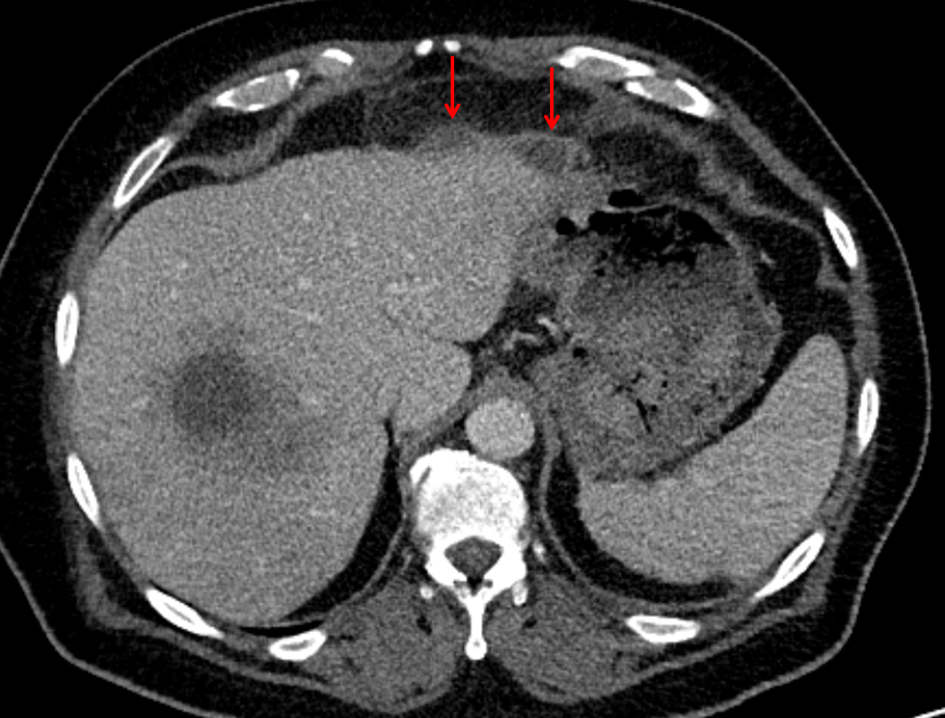
    • Large fluid density structure in hepatic segments 7 and 8 measuring 10 x 7 x 7 cm with internal septation and circumferential ill-defined low density compatible with edema
    • Peripherally enhancing subcapsular collections along the anterior margin of the left hepatic lobe measuring 3 x 1 cm and 2 x 1 cm
    • Clearly marginated fluid density structure in segment 7 and several other scattered tiny hypodensities, which likely represent cysts
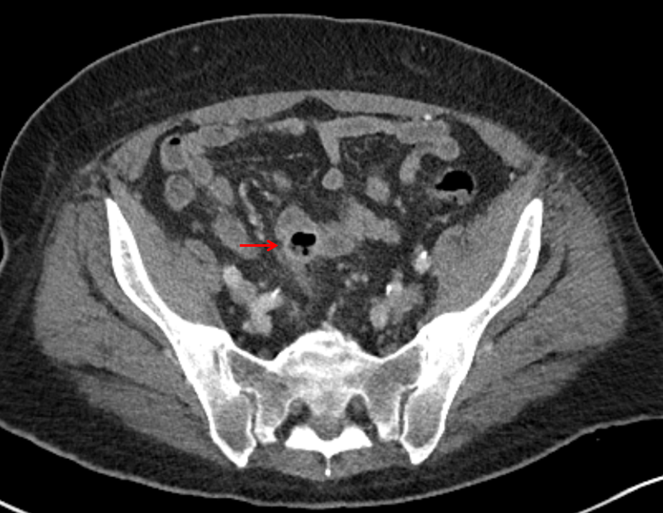
    • Mild mural thickening of a segment of the sigmoid colon with adjacent fat stranding and a 1.5 cm fluid and gas collection along the tip of an inflamed diverticulum
    • Loss of the normal fat plane between this collection and adjacent loops of small bowel, which demonstrate mural thickening
    • No bowel obstruction
    • High grade stenosis of the left common iliac artery, with the left internal and external iliac arteries remaining patent
    • Incidental splenule
  • MSK
    • No acute findings
    • Ankylosis of both sacroiliac joints
    • Circumscribed sclerotic lesion in the left anterolateral seventh rib without aggressive features, likely a bone island


Diagnosis

  • Hepatic abscess

Sample Report

Acute sigmoid diverticulitis complicated by a small contained perforation and a large abscess in the right hepatic lobe. Additional small subcapsular abscesses along the anterior margin of the left hepatic lobe.

Additionally, loss of the normal fat plane between the peridiverticular collection and adjacent thickened loops of small bowel raises the potential for an enterocolonic fistula.

High grade stenosis of the left common iliac artery. The left external and internal iliac arteries are patent.


Discussion

  • Pyogenic hepatic abscesses have many potential etiologies, including:
    • Direct spread from adjacent infection (e.g. gangrenous cholecystitis) or external introduction of infection (e.g. penetrating trauma)
    • Biliary spread from ascending cholangitis
    • Hematogenous spread (particularly from GI infections, as in this case)
  • The classic CT imaging appearance is a double target sign with internal low density surrounded by an internal enhancing rim (capsule) and a low density external rim (edema)
  • Abscesses may be unilocular or multilocular
  • Gas is present in a minority of cases
  • Unfortunately, necrotic tumors can have an overlapping imaging appearance, so it is important to follow these to resolution


Images

Hepatic abscess showing the double target sign with low density internally surrounded by a thin inner enhancing rim (red arrow) and ill-defined outer low density rim (yellow arrow). Blue arrow indicates an internal septation. Red arrows: additional smaller subcapsular abscesses. Red arrow: focal contained perforation associated with diverticulitis.


📣 Feedback?

⌨️  Keyboard Shortcuts ("K")

Call Preparation

Prepare for call efficiently with interactive cases, sample reports, and annotated images.

1,400+ cases  91.25 CME

Call Simulator

   Neuro CT

250 cases

  Neuro MRI

146 cases

   Peds Radiographs

372 cases

   MSK Radiographs

281 cases

   Chest CT

84 cases

   Chest Radiographs

97 cases

   KUB

43 cases

   Body CT

140 cases

   Ultrasound

75 cases

Call Simulator

• Shuffle cases from our courses to simulate the mix of a call shift.
• Submit your own report before reviewing the case write-up.

Neuro Fellowship

Reviews of neuro topics with clinical pearls, differentials, and in-depth discussions.

131 cases  13.1 CME

   Brain Tumors

105 cases

   Neurodegenerative

13 cases

   Congenital Hearing Loss

13 cases

Call Preparation

   Neuro CT

250 cases

   Neuro MRI

146 cases

   Peds Radiographs

372 cases

   MSK Radiographs

281 cases

   Ultrasound

75 cases

   Chest CT

84 cases

   Chest Radiographs

97 cases

   Body CT

127 cases

   KUB

43 cases

Neuro Fellowship

   Brain Tumors

105 cases

   Neurodegenerative

13 cases

Call Simulator

• Shuffle cases from our courses.
• Submit your own report.

CME

• Earn up to 91 CME.
• Claim your CME to receive a certificate.

Call Simulator

• Shuffle cases from our courses to simulate the mix of a call shift.
• Submit your own report before reviewing the case write-up.

CME

• Earn up to 91 CME by completing cases in our radiology courses.
• Claim your CME to receive a certificate.

On Call

Quick references created specifically to help while on call.

  Peds Normals by Age

Reference database of normal imaging from birth to age 16

   Incidental Findings

Summary of consensus guidelines for managing incidental CT findings

   Media Index

Index of select illustrations & videos from our courses

  Neuro CT Mimics

Visual reference for common mimics of pathology on CT

On Call

  Peds Normals by Age

Reference database of normal imaging from birth to age 16

   Incidental Findings

Summary of consensus guidelines for managing incidental CT findings

   Media Index

Index of select illustrations & videos from our courses

  Neuro CT Mimics

Visual reference for common mimics of pathology on CT

Anatomy

Labelled radiographs and CT/MRI series teaching anatomy with a level of detail appropriate for medical students and junior residents.

   Pelvis

Pelvic MRI anatomy

   Chest

Chest radiograph & CT anatomy

   Body

Abdominal CT anatomy

   Cardiac

Cardiac CT anatomy

   Brain

Brain & calvarial anatomy on CT/MRI

   Cranial Nerves

Cranial nerves on MRI

   Shoulder

Shoulder MRI anatomy

   Knee

Knee MRI anatomy

   Temporal Bone

Resident/fellow-level anatomy

Anatomy

   Pelvis

Pelvic MRI anatomy

   Chest

Chest radiograph & CT anatomy

   Body

Abdominal CT anatomy

   Cardiac

Cardiac CT anatomy

   Brain

Brain & calvarial anatomy on CT/MRI

   Cranial Nerves

Cranial nerves on MRI

   Shoulder

Shoulder MRI anatomy

   Knee

Knee MRI anatomy

   Temporal Bone

Resident/fellow-level anatomy

Neuro Courses

Includes our call preparation neuro courses (Neuro CT and Neuro MRI) and our neuro fellowship courses.

   Neuro CT

250 cases

  Neuro MRI

146 cases

   Brain Tumors

105 cases

   Neurodegenerative

13 cases

   Congenital Hearing Loss

Coming soon

Neuro Courses

   Neuro CT

250 cases

   Neuro MRI

146 cases

   Brain Tumors

105 cases

   Neurodegenerative

13 cases

  Congenital Hearing Loss

Coming soon

CME

• Claim your CME to receive a certificate.

Call Simulator

• Shuffle cases from our courses to simulate the mix of a call shift.
• Submit your own report before reviewing the case write-up.

CME

• Earn up to 91 CME by completing cases in our radiology courses.
• Claim your CME to receive a certificate.

   Neuro CT

250 cases

   Neuro MRI

146 cases

   Peds Radiographs

372 cases

   MSK Radiographs

281 cases

   Ultrasound

75 cases

   Chest CT

84 cases

   Chest Radiographs

97 cases

   Body CT

140 cases

   KUB

43 cases

Call Simulator

• Shuffle cases from our courses to simulate the mix of a call shift.
• Submit your own report before reviewing the case write-up.

CME

• Earn up to 91 CME by completing cases in our radiology courses.
• Claim your CME to receive a certificate.

⌨️ Keyboard Shortcuts

View shortcuts

Zoom/Pan

  Full screen

  Window/Level

  Expand/collapse

  Scroll

  Save the case

  Close case/tab

Previous Slice (PC)

Next Slice (PC)

SHIFT Previous Slice (MAC)

 SHIFT Next Slice (MAC)

 Toggle presets

 Previous series (if multiple)

 Next series (if multiple)

  Previous case (⌘ ◄ on a mac)

  Next case (⌘ ► on a mac)

Toggle dark mode