Expand ("E")   Collapse

Age: 75

Sex: Male

Indication: Fall

Save ("V")

Case #3


Findings

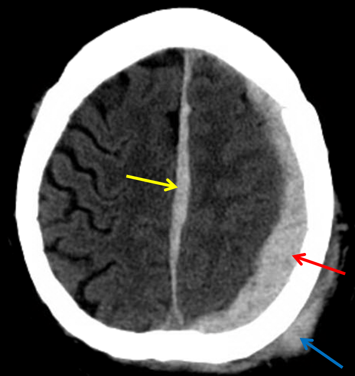
  • Acute subdural hematoma layering over the left cerebral convexity with a smaller portion extending over the left tentorium and interhemispheric falx
  • Diffuse left cerebral hemispheric sulcal effacement with left to right midline shift
  • Left posterior scalp hematoma
  • Right medial orbital wall blow-out fracture without hemorrhage in the adjacent ethmoid air cells or extraconal fat. No extraocular muscle herniation
  • Incidental note is made of dolichoectasia of the vertebral and basilar arteries


Diagnosis

  • Acute subdural hematoma

Sample Report

Acute subdural hematoma measuring up to 10 mm in thickness layering over the left cerebral convexity with a smaller portion extending over the tentorium and interhemispheric falx. This results in diffuse left cerebral hemispheric sulcal effacement with 6 mm left to right midline shift. Basal cisterns are patent without evidence of uncal or cerebellar tonsillar herniation.

Left posterior scalp hematoma without subjacent calvarial fracture.

Right medial orbital wall blow-out fracture without hemorrhage in the adjacent ethmoid air cells or extraconal fat, favoring a remote fracture. No extraocular muscle herniation.


Discussion

  • Subdural hematomas accumulate between the dura and arachnoid mater
  • These are seen more commonly than epidural hematomas and often occur in the elderly, though they are also seen with nonaccidental trauma in infants and in trauma across all ages
  • Subdural hematomas result from venous bleeding, typically due to rupture of bridging cortical veins which traverse the potential subdural space on their way to the dural venous sinuses
  • There is a less common association with skull fractures
  • Though not always easy to tell apart from epidural hematomas, favor subdural hematoma if:
    • A hematoma crosses suture lines but respects midline (epidural hematomas respect suture lines unless there is a fracture through a suture or sutural diastasis)
    • A hematoma is crescent-shaped rather than lens-shaped and layers along the entire cerebral convexity or extends along the falx or tentorium
  • Here is the typical progression of density of blood products on noncontrast CT (*please note that these are estimates that can vary from case to case and can be substantially altered in the setting of coagulopathy; also if the patient is anemic, even acute blood may not be very dense appearing*)
    • Hyperacute (< 1 hr) – hypodense (unclotted blood)
    • Acute (hours to weeks) – hyperdense
    • Subacute (~2-4 weeks) – isodense to cortex – these are really tough to see
    • Chronic (months) – hypodense to cortex and eventually isodense to CSF
  • Always mention fractures even if you are pretty sure they are old (like the medial orbital wall fracture in this case). Sometimes acute fractures can fool you by not having much surrounding edema/hemorrhage
  • Scalp contusions and hematomas often go together. Although not a strict rule, contusions are typically less well defined (these represent edema secondary to tissue damage and often cause subcutaneous fat stranding) and hematomas are often more hyperattenuating and demarcated


Images

Red arrow: acute subdural hematoma layering along the left cerebral convexity. Yellow arrow: subdural hematoma layering along the interhemispheric falx. Blue arrow: scalp hematoma.

Video Reviews

Evolution of Hemorrhage on Head CT


📣 Feedback?

⌨️  Keyboard Shortcuts ("K")

Call Preparation

Prepare for call efficiently with interactive cases, sample reports, and annotated images.

1,400+ cases  91.25 CME

Call Simulator

   Neuro CT

250 cases

  Neuro MRI

146 cases

   Peds Radiographs

372 cases

   MSK Radiographs

281 cases

   Chest CT

84 cases

   Chest Radiographs

97 cases

   KUB

43 cases

   Body CT

140 cases

   Ultrasound

75 cases

Call Simulator

• Shuffle cases from our courses to simulate the mix of a call shift.
• Submit your own report before reviewing the case write-up.

Neuro Fellowship

Reviews of neuro topics with clinical pearls, differentials, and in-depth discussions.

131 cases  13.1 CME

   Brain Tumors

105 cases

   Neurodegenerative

13 cases

   Congenital Hearing Loss

13 cases

Call Preparation

   Neuro CT

250 cases

   Neuro MRI

146 cases

   Peds Radiographs

372 cases

   MSK Radiographs

281 cases

   Ultrasound

75 cases

   Chest CT

84 cases

   Chest Radiographs

97 cases

   Body CT

127 cases

   KUB

43 cases

Neuro Fellowship

   Brain Tumors

105 cases

   Neurodegenerative

13 cases

Call Simulator

• Shuffle cases from our courses.
• Submit your own report.

CME

• Earn up to 91 CME.
• Claim your CME to receive a certificate.

Call Simulator

• Shuffle cases from our courses to simulate the mix of a call shift.
• Submit your own report before reviewing the case write-up.

CME

• Earn up to 91 CME by completing cases in our radiology courses.
• Claim your CME to receive a certificate.

On Call

Quick references created specifically to help while on call.

  Peds Normals by Age

Reference database of normal imaging from birth to age 16

   Incidental Findings

Summary of consensus guidelines for managing incidental CT findings

   Media Index

Index of select illustrations & videos from our courses

  Neuro CT Mimics

Visual reference for common mimics of pathology on CT

On Call

  Peds Normals by Age

Reference database of normal imaging from birth to age 16

   Incidental Findings

Summary of consensus guidelines for managing incidental CT findings

   Media Index

Index of select illustrations & videos from our courses

  Neuro CT Mimics

Visual reference for common mimics of pathology on CT

Anatomy

Labelled radiographs and CT/MRI series teaching anatomy with a level of detail appropriate for medical students and junior residents.

   Pelvis

Pelvic MRI anatomy

   Chest

Chest radiograph & CT anatomy

   Body

Abdominal CT anatomy

   Cardiac

Cardiac CT anatomy

   Brain

Brain & calvarial anatomy on CT/MRI

   Cranial Nerves

Cranial nerves on MRI

   Shoulder

Shoulder MRI anatomy

   Knee

Knee MRI anatomy

   Temporal Bone

Resident/fellow-level anatomy

Anatomy

   Pelvis

Pelvic MRI anatomy

   Chest

Chest radiograph & CT anatomy

   Body

Abdominal CT anatomy

   Cardiac

Cardiac CT anatomy

   Brain

Brain & calvarial anatomy on CT/MRI

   Cranial Nerves

Cranial nerves on MRI

   Shoulder

Shoulder MRI anatomy

   Knee

Knee MRI anatomy

   Temporal Bone

Resident/fellow-level anatomy

Neuro Courses

Includes our call preparation neuro courses (Neuro CT and Neuro MRI) and our neuro fellowship courses.

   Neuro CT

250 cases

  Neuro MRI

146 cases

   Brain Tumors

105 cases

   Neurodegenerative

13 cases

   Congenital Hearing Loss

Coming soon

Neuro Courses

   Neuro CT

250 cases

   Neuro MRI

146 cases

   Brain Tumors

105 cases

   Neurodegenerative

13 cases

  Congenital Hearing Loss

Coming soon

CME

• Claim your CME to receive a certificate.

Call Simulator

• Shuffle cases from our courses to simulate the mix of a call shift.
• Submit your own report before reviewing the case write-up.

CME

• Earn up to 91 CME by completing cases in our radiology courses.
• Claim your CME to receive a certificate.

   Neuro CT

250 cases

   Neuro MRI

146 cases

   Peds Radiographs

372 cases

   MSK Radiographs

281 cases

   Ultrasound

75 cases

   Chest CT

84 cases

   Chest Radiographs

97 cases

   Body CT

140 cases

   KUB

43 cases

Call Simulator

• Shuffle cases from our courses to simulate the mix of a call shift.
• Submit your own report before reviewing the case write-up.

CME

• Earn up to 91 CME by completing cases in our radiology courses.
• Claim your CME to receive a certificate.

⌨️ Keyboard Shortcuts

View shortcuts

Zoom/Pan

  Full screen

  Window/Level

  Expand/collapse

  Scroll

  Save the case

  Close case/tab

Previous Slice (PC)

Next Slice (PC)

SHIFT Previous Slice (MAC)

 SHIFT Next Slice (MAC)

 Toggle presets

 Previous series (if multiple)

 Next series (if multiple)

  Previous case (⌘ ◄ on a mac)

  Next case (⌘ ► on a mac)

Toggle dark mode