Become a Pro Member for full access to this course and more.

View Plans   Log In
Expand ("E")   Collapse

Age: 46

Sex: Male

Indication: Trauma

Save ("V")

Case #20


Findings

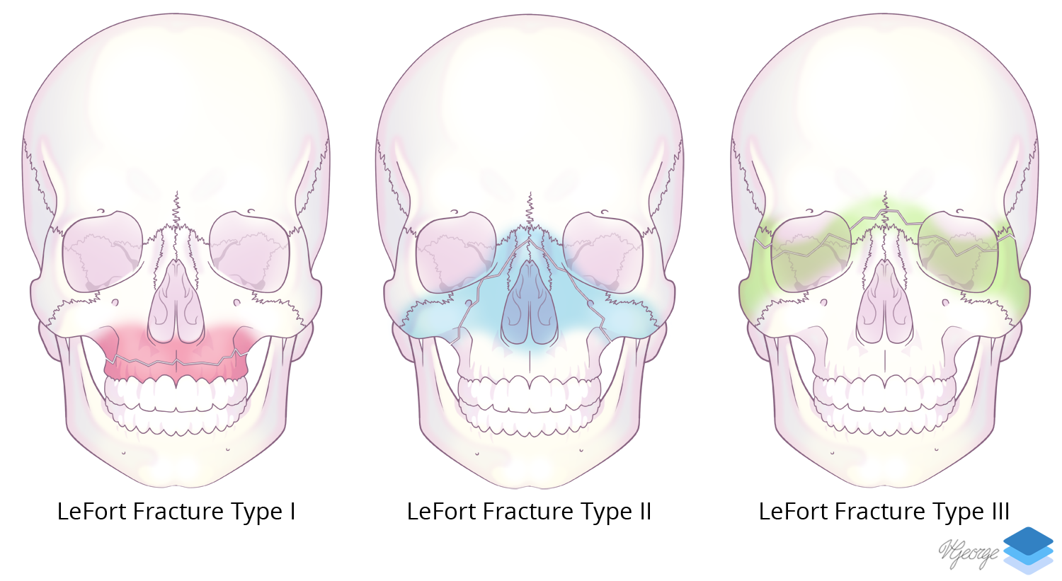
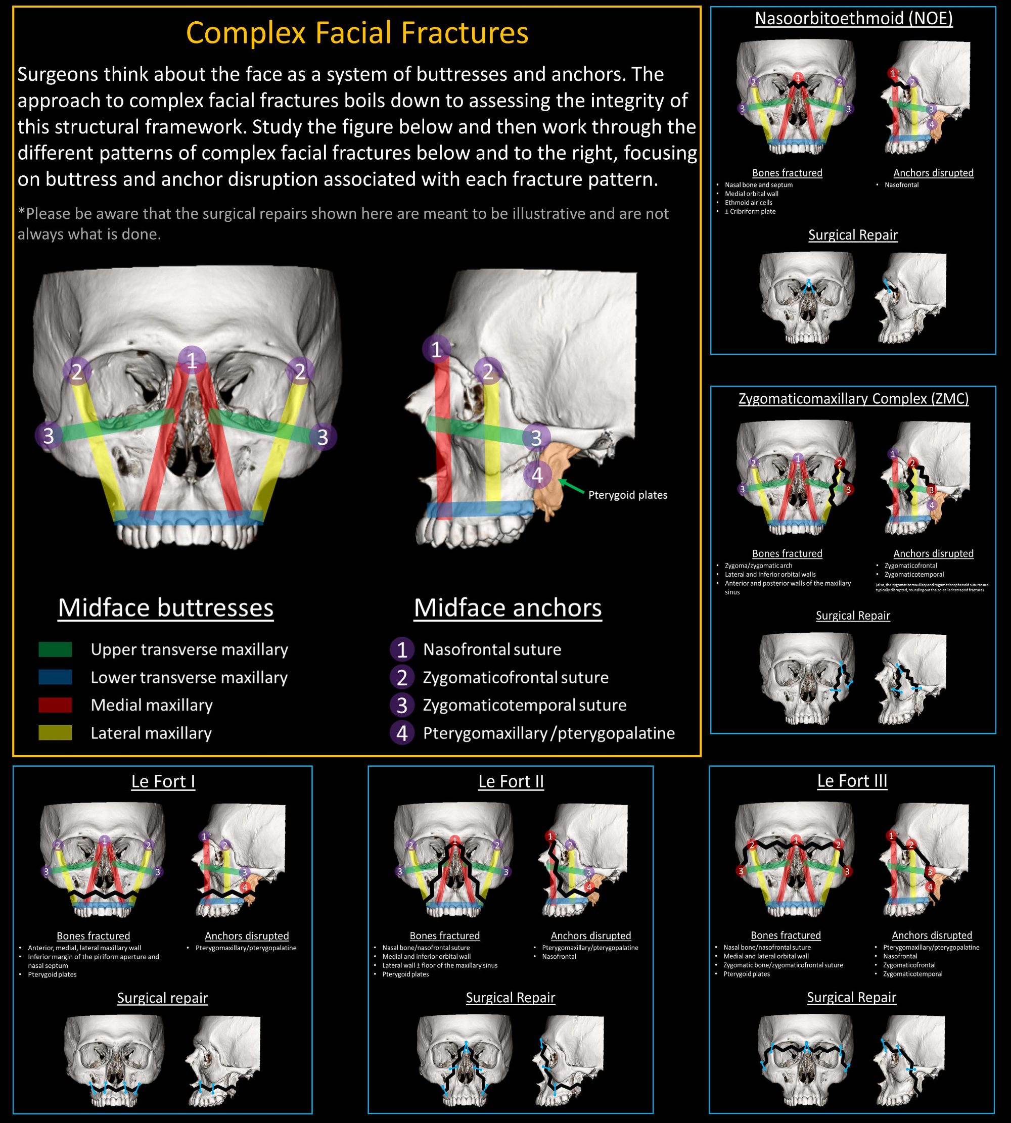
  • Right Le Fort type I, II, and III, zygomaticomaxillary complex (ZMC), and nasoorbitoethmoid (NOE) fractures with a comminuted fracture of the right zygomatic arch, blowout fractures of the right medial, lateral, and inferior orbital walls, fractures of all walls of the right maxillary sinus, bilateral nasal arch fractures, fractures throughout the right ethmoid air cells extending superiorly through the cribriform plate, fracture of the nasal septum, and fractures of the right pterygoid plates. Fractures involve the nasolacrimal canal and infraorbital foramen
  • Herniation of orbital and retroantral fat through the right orbital floor blowout fracture and posterior maxillary wall fracture, respectively,  with contour irregularity of the right medial and inferior rectus muscles. Right retrobulbar hemorrhage and gas without proptosis
  • Acute depressed right parietal fracture with anterior continuation as a comminuted fracture of the greater wing of the right sphenoid with a comminuted fracture of the lateral wall of the right sphenoid sinus extending to involve the right carotid canal, right foramen rotundum, and right inferior orbital foramen. There is also continuation through the right frontal bone with involvement of the frontal sinus and into the lateral wall and roof of the right orbit
  • Acute nondisplaced left parietal fracture extending into the left frontal bone, involving the left frontal sinus and extending into the lateral wall and roof of the left orbit. Left retrobulbar hemorrhage and gas without proptosis
  •  Bilateral diastasis of the zygomaticofrontal sutures
  • Acute minimally displaced fracture of the right hemimandible at the base of the coronoid process extending into the condylar neck
  • Bifrontal extraaxial hematomas and pneumocephalus
  • Extensive hemosinus
  • Extensive facial soft tissue swelling and subcutaneous emphysema


Diagnosis

  • Extensive facial smash injury, skull fractures, extraaxial hematomas

Sample Report

Extensive facial smash injury with right Le Fort type I, II, and III, zygomaticomaxillary complex (ZMC), and nasoorbitoethmoid (NOE) fractures. Notably, fractures involve the nasolacrimal canal and infraorbital foramen and extend superiorly through the cribriform plate on the right. There is bilateral diastasis of the zygomaticofrontal sutures.

Partially imaged acute depressed right parietal fracture with anterior continuation as a comminuted fracture of the greater wing of the right sphenoid with a comminuted fracture of the lateral wall of the right sphenoid sinus extending to involve the right carotid canal, right foramen rotundum, and right inferior orbital foramen. There is also continuation through the right frontal bone with involvement of the frontal sinus and into the lateral wall and roof of the right orbit. Additionally, there is an acute nondisplaced left parietal fracture extending into the left frontal bone, involving the left frontal sinus and extending into the lateral wall and roof of the left orbit. Bifrontal extraaxial hematomas and pneumocephalus, which can be further assessed on dedicated head CT imaging.

Herniation of orbital and retroantral fat through a right orbital floor blowout fracture and posterior maxillary wall fracture, respectively,  with contour irregularity of the right medial and inferior rectus muscles. Recommend correlation with clinical signs of extraocular muscle entrapment.

Bilateral retrobulbar hemorrhage and gas without proptosis. Globes are intact.

Acute minimally displaced fracture of the right hemimandible at the base of the coronoid process extending into the condylar neck. Temporomandibular joints remain located.

Extensive hemosinus.

Extensive facial soft tissue swelling and subcutaneous emphysema.


Discussion

Become a Pro Member to unlock the discussion.

View Plans   Log In


Images

LeFort Fracture Classification Complex facial fracture classification, including Le Fort I, II, and III. Full size image at: bit.ly/leforts


📣 Feedback?

⌨️  Keyboard Shortcuts ("K")

Call Preparation

Prepare for call efficiently with interactive cases, sample reports, and annotated images.

1,400+ cases  91.25 CME

Call Simulator

   Neuro CT

250 cases

  Neuro MRI

146 cases

   Peds Radiographs

372 cases

   MSK Radiographs

281 cases

   Chest CT

84 cases

   Chest Radiographs

97 cases

   KUB

43 cases

   Body CT

140 cases

   Ultrasound

75 cases

Call Simulator

• Shuffle cases from our courses to simulate the mix of a call shift.
• Submit your own report before reviewing the case write-up.

Neuro Fellowship

Reviews of neuro topics with clinical pearls, differentials, and in-depth discussions.

131 cases  13.1 CME

   Brain Tumors

105 cases

   Neurodegenerative

13 cases

   Congenital Hearing Loss

13 cases

Call Preparation

   Neuro CT

250 cases

   Neuro MRI

146 cases

   Peds Radiographs

372 cases

   MSK Radiographs

281 cases

   Ultrasound

75 cases

   Chest CT

84 cases

   Chest Radiographs

97 cases

   Body CT

127 cases

   KUB

43 cases

Neuro Fellowship

   Brain Tumors

105 cases

   Neurodegenerative

13 cases

Call Simulator

• Shuffle cases from our courses.
• Submit your own report.

CME

• Earn up to 91 CME.
• Claim your CME to receive a certificate.

Call Simulator

• Shuffle cases from our courses to simulate the mix of a call shift.
• Submit your own report before reviewing the case write-up.

CME

• Earn up to 91 CME by completing cases in our radiology courses.
• Claim your CME to receive a certificate.

On Call

Quick references created specifically to help while on call.

  Peds Normals by Age

Reference database of normal imaging from birth to age 16

   Incidental Findings

Summary of consensus guidelines for managing incidental CT findings

   Media Index

Index of select illustrations & videos from our courses

  Neuro CT Mimics

Visual reference for common mimics of pathology on CT

On Call

  Peds Normals by Age

Reference database of normal imaging from birth to age 16

   Incidental Findings

Summary of consensus guidelines for managing incidental CT findings

   Media Index

Index of select illustrations & videos from our courses

  Neuro CT Mimics

Visual reference for common mimics of pathology on CT

Anatomy

Labelled radiographs and CT/MRI series teaching anatomy with a level of detail appropriate for medical students and junior residents.

   Pelvis

Pelvic MRI anatomy

   Chest

Chest radiograph & CT anatomy

   Body

Abdominal CT anatomy

   Cardiac

Cardiac CT anatomy

   Brain

Brain & calvarial anatomy on CT/MRI

   Cranial Nerves

Cranial nerves on MRI

   Shoulder

Shoulder MRI anatomy

   Knee

Knee MRI anatomy

   Temporal Bone

Resident/fellow-level anatomy

Anatomy

   Pelvis

Pelvic MRI anatomy

   Chest

Chest radiograph & CT anatomy

   Body

Abdominal CT anatomy

   Cardiac

Cardiac CT anatomy

   Brain

Brain & calvarial anatomy on CT/MRI

   Cranial Nerves

Cranial nerves on MRI

   Shoulder

Shoulder MRI anatomy

   Knee

Knee MRI anatomy

   Temporal Bone

Resident/fellow-level anatomy

Neuro Courses

Includes our call preparation neuro courses (Neuro CT and Neuro MRI) and our neuro fellowship courses.

   Neuro CT

250 cases

  Neuro MRI

146 cases

   Brain Tumors

105 cases

   Neurodegenerative

13 cases

   Congenital Hearing Loss

Coming soon

Neuro Courses

   Neuro CT

250 cases

   Neuro MRI

146 cases

   Brain Tumors

105 cases

   Neurodegenerative

13 cases

  Congenital Hearing Loss

Coming soon

CME

• Claim your CME to receive a certificate.

Call Simulator

• Shuffle cases from our courses to simulate the mix of a call shift.
• Submit your own report before reviewing the case write-up.

CME

• Earn up to 91 CME by completing cases in our radiology courses.
• Claim your CME to receive a certificate.

   Neuro CT

250 cases

   Neuro MRI

146 cases

   Peds Radiographs

372 cases

   MSK Radiographs

281 cases

   Ultrasound

75 cases

   Chest CT

84 cases

   Chest Radiographs

97 cases

   Body CT

140 cases

   KUB

43 cases

Call Simulator

• Shuffle cases from our courses to simulate the mix of a call shift.
• Submit your own report before reviewing the case write-up.

CME

• Earn up to 91 CME by completing cases in our radiology courses.
• Claim your CME to receive a certificate.

⌨️ Keyboard Shortcuts

View shortcuts

Zoom/Pan

  Full screen

  Window/Level

  Expand/collapse

  Scroll

  Save the case

  Close case/tab

Previous Slice (PC)

Next Slice (PC)

SHIFT Previous Slice (MAC)

 SHIFT Next Slice (MAC)

 Toggle presets

 Previous series (if multiple)

 Next series (if multiple)

  Previous case (⌘ ◄ on a mac)

  Next case (⌘ ► on a mac)

Toggle dark mode