Expand ("E")   Collapse

Age: 5

Sex: Female

Indication: Headache, somnolence

Save ("V")

Case #1


Findings for this case

CT

  • Hypoattenuating mass centered in the fourth ventricle with a more solid appearing component superiorly
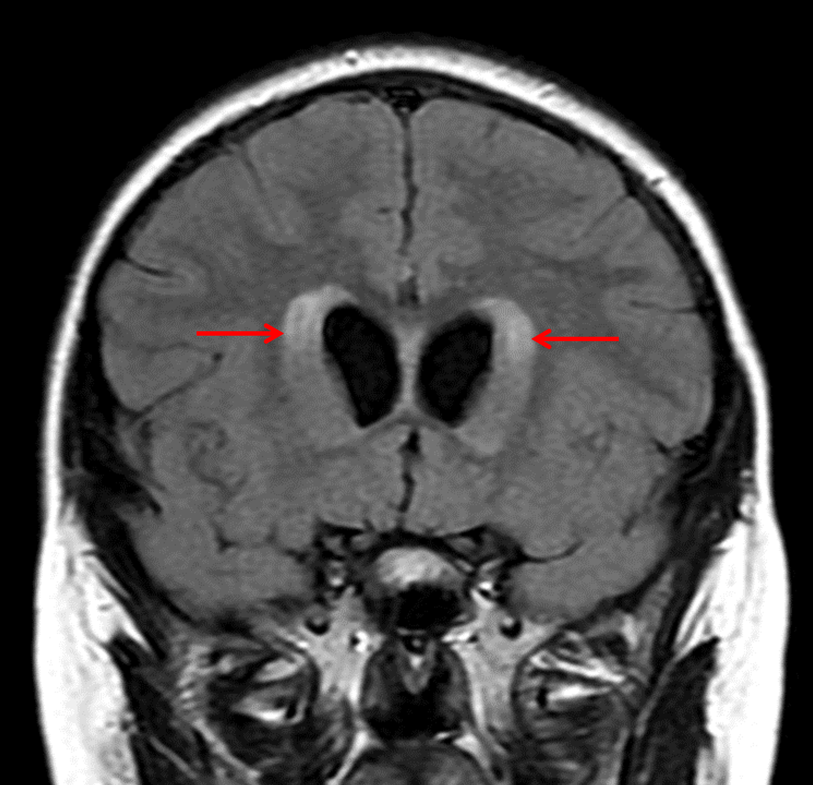
  • Associated obstructive hydrocephalus and periventricular edema


MRI

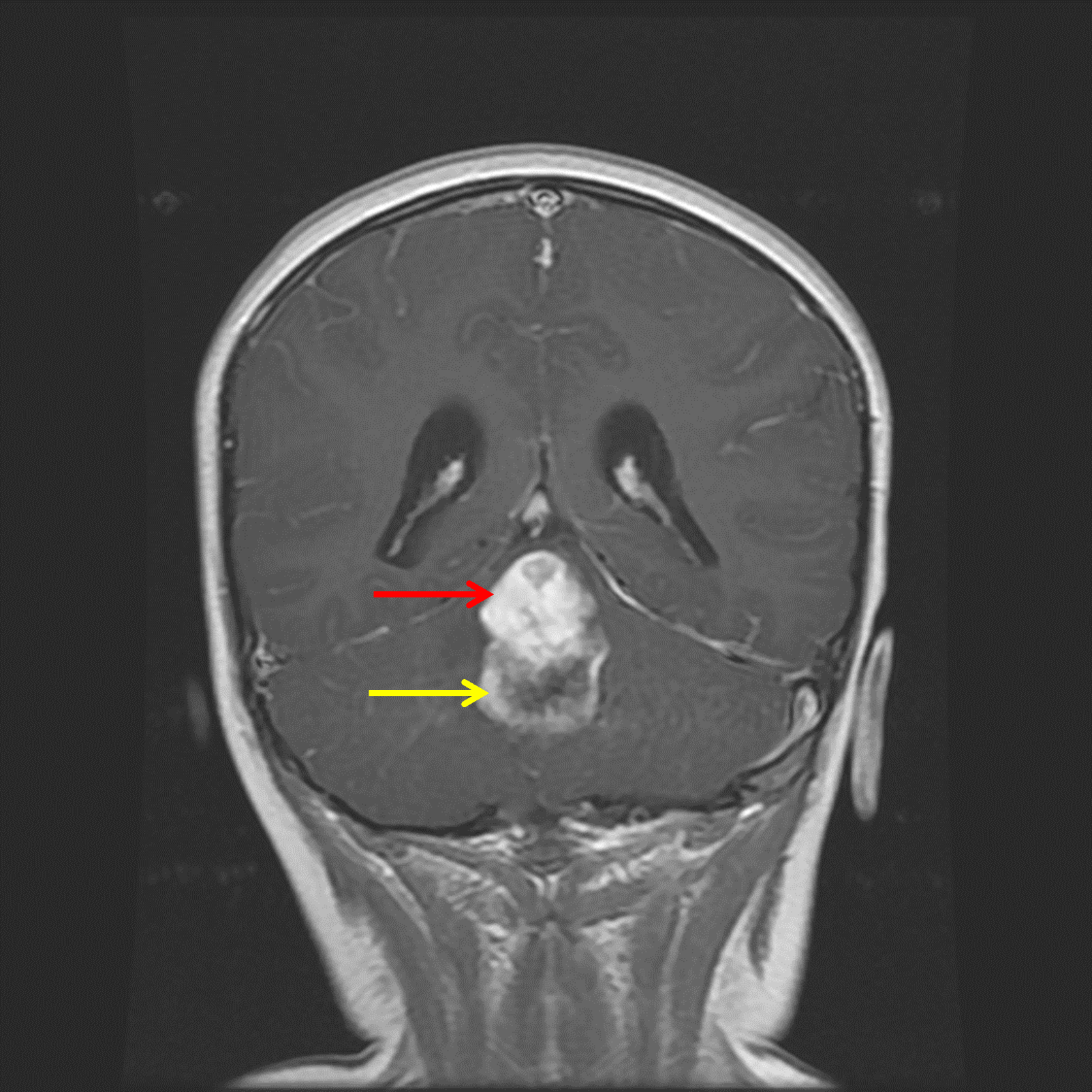
  • Mixed solid and cystic mass centered in the fourth ventricle measuring 2.5 x 2.5 x 4 cm with a more solid enhancing component superiorly and a peripherally-enhancing cystic component inferiorly
  • No corresponding restricted diffusion
  • Associated effacement of the fourth ventricle and obstructive hydrocephalus with periventricular edema


Images

Midline posterior fossa mass with a superior solid component (red arrow) and inferior peripherally-enhancing cystic component (yellow arrow). Associated obstructive hydrocephalus with subependymal edema (red arrows).

Diagnosis & Discussion

Diagnosis:

Pilocytic astrocytoma


Background

  • Think about JPA when you encounter a “cyst with nodule” mass, or any enhancing mass for that matter, in the posterior fossa in a child
  • There is a strong association between optic pathway gliomas and NF1

Imaging Findings

  • The “cyst with nodule” appearance is classic, but a minority of tumors do not demonstrate cystic components
  • The solid component is usually hypoattenuating to brain on CT, T2 hyperintense, solidly enhancing, and does not demonstrate restricted diffusion
  • Internal hemorrhage is rare
  • Calcification is not usually seen


Differential

  • Ependymoma – Avidly enhancing tumors with a predilection for the fourth ventricle in children, these tend to be more malleable (including the characteristic appearance of tumor bulging through the foramina of Luschka) and are less likely to have cystic components
  • Medulloblastoma – Avidly enhancing, commonly midline posterior fossa tumors, these tend to present at a younger age (3-9 years old) and classically have associated restricted diffusion
  • Hemangioblastoma – Also “cyst with nodule” tumors, these are more common in adults and in patients with von Hippel Lindau disease
  • High-grade glioma – These tend to occur in older children and are often more heterogeneous and poorly marginated than JPAs. Also, unlike JPAs, these have a propensity for CSF dissemination


Pearls

  • Think about JPA when you encounter a “cyst with nodule” mass, or any enhancing mass for that matter, in the posterior fossa in a child
  • There is a strong association between optic pathway gliomas and NF1


📣 Feedback?

⌨️  Keyboard Shortcuts ("K")

Call Preparation

Prepare for call efficiently with interactive cases, sample reports, and annotated images.

1,400+ cases  91.25 CME

Call Simulator

   Neuro CT

250 cases

  Neuro MRI

146 cases

   Peds Radiographs

372 cases

   MSK Radiographs

281 cases

   Chest CT

84 cases

   Chest Radiographs

97 cases

   KUB

43 cases

   Body CT

140 cases

   Ultrasound

75 cases

Call Simulator

• Shuffle cases from our courses to simulate the mix of a call shift.
• Submit your own report before reviewing the case write-up.

Neuro Fellowship

Reviews of neuro topics with clinical pearls, differentials, and in-depth discussions.

131 cases  13.1 CME

   Brain Tumors

105 cases

   Neurodegenerative

13 cases

   Congenital Hearing Loss

13 cases

Call Preparation

   Neuro CT

250 cases

   Neuro MRI

146 cases

   Peds Radiographs

372 cases

   MSK Radiographs

281 cases

   Ultrasound

75 cases

   Chest CT

84 cases

   Chest Radiographs

97 cases

   Body CT

127 cases

   KUB

43 cases

Neuro Fellowship

   Brain Tumors

105 cases

   Neurodegenerative

13 cases

Call Simulator

• Shuffle cases from our courses.
• Submit your own report.

CME

• Earn up to 91 CME.
• Claim your CME to receive a certificate.

Call Simulator

• Shuffle cases from our courses to simulate the mix of a call shift.
• Submit your own report before reviewing the case write-up.

CME

• Earn up to 91 CME by completing cases in our radiology courses.
• Claim your CME to receive a certificate.

On Call

Quick references created specifically to help while on call.

  Peds Normals by Age

Reference database of normal imaging from birth to age 16

   Incidental Findings

Summary of consensus guidelines for managing incidental CT findings

   Media Index

Index of select illustrations & videos from our courses

  Neuro CT Mimics

Visual reference for common mimics of pathology on CT

On Call

  Peds Normals by Age

Reference database of normal imaging from birth to age 16

   Incidental Findings

Summary of consensus guidelines for managing incidental CT findings

   Media Index

Index of select illustrations & videos from our courses

  Neuro CT Mimics

Visual reference for common mimics of pathology on CT

Anatomy

Labelled radiographs and CT/MRI series teaching anatomy with a level of detail appropriate for medical students and junior residents.

   Pelvis

Pelvic MRI anatomy

   Chest

Chest radiograph & CT anatomy

   Body

Abdominal CT anatomy

   Cardiac

Cardiac CT anatomy

   Brain

Brain & calvarial anatomy on CT/MRI

   Cranial Nerves

Cranial nerves on MRI

   Shoulder

Shoulder MRI anatomy

   Knee

Knee MRI anatomy

   Temporal Bone

Resident/fellow-level anatomy

Anatomy

   Pelvis

Pelvic MRI anatomy

   Chest

Chest radiograph & CT anatomy

   Body

Abdominal CT anatomy

   Cardiac

Cardiac CT anatomy

   Brain

Brain & calvarial anatomy on CT/MRI

   Cranial Nerves

Cranial nerves on MRI

   Shoulder

Shoulder MRI anatomy

   Knee

Knee MRI anatomy

   Temporal Bone

Resident/fellow-level anatomy

Neuro Courses

Includes our call preparation neuro courses (Neuro CT and Neuro MRI) and our neuro fellowship courses.

   Neuro CT

250 cases

  Neuro MRI

146 cases

   Brain Tumors

105 cases

   Neurodegenerative

13 cases

   Congenital Hearing Loss

Coming soon

Neuro Courses

   Neuro CT

250 cases

   Neuro MRI

146 cases

   Brain Tumors

105 cases

   Neurodegenerative

13 cases

  Congenital Hearing Loss

Coming soon

CME

• Claim your CME to receive a certificate.

Call Simulator

• Shuffle cases from our courses to simulate the mix of a call shift.
• Submit your own report before reviewing the case write-up.

CME

• Earn up to 91 CME by completing cases in our radiology courses.
• Claim your CME to receive a certificate.

   Neuro CT

250 cases

   Neuro MRI

146 cases

   Peds Radiographs

372 cases

   MSK Radiographs

281 cases

   Ultrasound

75 cases

   Chest CT

84 cases

   Chest Radiographs

97 cases

   Body CT

140 cases

   KUB

43 cases

Call Simulator

• Shuffle cases from our courses to simulate the mix of a call shift.
• Submit your own report before reviewing the case write-up.

CME

• Earn up to 91 CME by completing cases in our radiology courses.
• Claim your CME to receive a certificate.

⌨️ Keyboard Shortcuts

View shortcuts

Zoom/Pan

  Full screen

  Window/Level

  Expand/collapse

  Scroll

  Save the case

  Close case/tab

Previous Slice (PC)

Next Slice (PC)

SHIFT Previous Slice (MAC)

 SHIFT Next Slice (MAC)

 Toggle presets

 Previous series (if multiple)

 Next series (if multiple)

  Previous case (⌘ ◄ on a mac)

  Next case (⌘ ► on a mac)

Toggle dark mode