Expand ("E")   Collapse

Age: 75

Sex: Male

Indication: Dementia

Save ("V")

Case #1


Findings

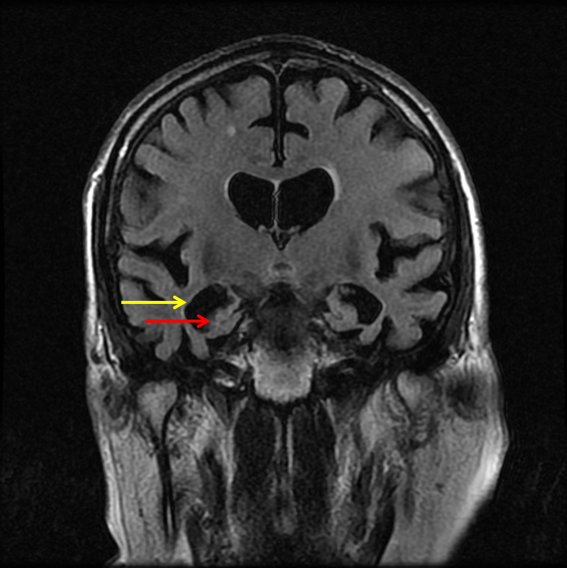
  • Symmetric volume loss involving both temporal lobes and hippocampi with ex vacuo enlargement of the temporal horns of both lateral ventricles, which is out of proportion to the degree of generalized cerebral and cerebellar volume loss
  • Patchy T2/FLAIR hyperintensity in the subcortical and periventricular white matter, which although nonspecific likely relates to chronic small vessel disease
  • T2 hyperintense lesion extending from the posterior aspect of the right nasal cavity into the nasopharynx, likely a polyp
  • Bilateral ethmoid air cell mucosal thickening
  • Bilateral pseudophakia
  • Anterolisthesis at C3-C4 results in mild spinal canal narrowing


Diagnosis

Alzheimer disease

Sample Report

Advanced bilateral temporal and hippocampal volume loss out of proportion to the over degree of cerebral atrophy, which can be seen with Alzheimer disease.

No acute intracranial abnormality.


Discussion

  • Clinical
    • Alzheimer disease is the most common cause of dementia in the elderly
    • Progressive cognitive impairment (often beginning with memory loss followed by apraxia and loss of executive function) that gradually leads to loss of functional independence
    • Patients may survive 10 years or more after diagnosis
  • Histopathology/Genetics
    • Accumulation of beta amyloid plaques in cortical gray matter and in arteriole walls
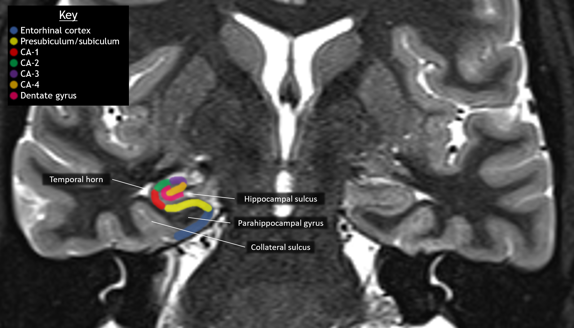
    • Abnormal tau phosphorylation leads to development of neurofibrillary tangles (seen earliest in the entorhinal cortex)
    • A minority of cases are familial arising in the setting of genetic mutations in several genes including the amyloid precursor protein (APP) gene
  • Imaging Findings
    • Atrophy (and corresponding hypometabolism on PET imaging) in the following areas:
      • Mesial temporal lobes including the entorhinal cortex and hippocampus (especially CA-1) – look for disproportionate ex vacuo enlargement of the temporal horns
      • Posterior cingulate gyri
      • Parietal lobes (especially precuneus), sparing the primary cortex until late in the disease
    • May see findings of amyloid angiopathy (multiple lobar microhemorrhages relatively sparing the deep gray structures)
    • Amyloid-specific PET agents show abnormal cerebral gray matter radiotracer uptake


Images

Anatomy of the mesial temporal structures on a coronal STIR image from a young, healthy patient. Disproportionate volume loss of the hippocampi and parahippocampal gyri (red arrow) with ex vacuo temporal horn enlargement (yellow arrow) in this patient with Alzheimer disease.


📣 Feedback?

⌨️  Keyboard Shortcuts ("K")

Call Preparation

Prepare for call efficiently with interactive cases, sample reports, and annotated images.

1,400+ cases  91.25 CME

Call Simulator

   Neuro CT

250 cases

  Neuro MRI

146 cases

   Peds Radiographs

372 cases

   MSK Radiographs

281 cases

   Chest CT

84 cases

   Chest Radiographs

97 cases

   KUB

43 cases

   Body CT

140 cases

   Ultrasound

75 cases

Call Simulator

• Shuffle cases from our courses to simulate the mix of a call shift.
• Submit your own report before reviewing the case write-up.

Neuro Fellowship

Reviews of neuro topics with clinical pearls, differentials, and in-depth discussions.

131 cases  13.1 CME

   Brain Tumors

105 cases

   Neurodegenerative

13 cases

   Congenital Hearing Loss

13 cases

Call Preparation

   Neuro CT

250 cases

   Neuro MRI

146 cases

   Peds Radiographs

372 cases

   MSK Radiographs

281 cases

   Ultrasound

75 cases

   Chest CT

84 cases

   Chest Radiographs

97 cases

   Body CT

127 cases

   KUB

43 cases

Neuro Fellowship

   Brain Tumors

105 cases

   Neurodegenerative

13 cases

Call Simulator

• Shuffle cases from our courses.
• Submit your own report.

CME

• Earn up to 91 CME.
• Claim your CME to receive a certificate.

Call Simulator

• Shuffle cases from our courses to simulate the mix of a call shift.
• Submit your own report before reviewing the case write-up.

CME

• Earn up to 91 CME by completing cases in our radiology courses.
• Claim your CME to receive a certificate.

On Call

Quick references created specifically to help while on call.

  Peds Normals by Age

Reference database of normal imaging from birth to age 16

   Incidental Findings

Summary of consensus guidelines for managing incidental CT findings

   Media Index

Index of select illustrations & videos from our courses

  Neuro CT Mimics

Visual reference for common mimics of pathology on CT

On Call

  Peds Normals by Age

Reference database of normal imaging from birth to age 16

   Incidental Findings

Summary of consensus guidelines for managing incidental CT findings

   Media Index

Index of select illustrations & videos from our courses

  Neuro CT Mimics

Visual reference for common mimics of pathology on CT

Anatomy

Labelled radiographs and CT/MRI series teaching anatomy with a level of detail appropriate for medical students and junior residents.

   Pelvis

Pelvic MRI anatomy

   Chest

Chest radiograph & CT anatomy

   Body

Abdominal CT anatomy

   Cardiac

Cardiac CT anatomy

   Brain

Brain & calvarial anatomy on CT/MRI

   Cranial Nerves

Cranial nerves on MRI

   Shoulder

Shoulder MRI anatomy

   Knee

Knee MRI anatomy

   Temporal Bone

Resident/fellow-level anatomy

Anatomy

   Pelvis

Pelvic MRI anatomy

   Chest

Chest radiograph & CT anatomy

   Body

Abdominal CT anatomy

   Cardiac

Cardiac CT anatomy

   Brain

Brain & calvarial anatomy on CT/MRI

   Cranial Nerves

Cranial nerves on MRI

   Shoulder

Shoulder MRI anatomy

   Knee

Knee MRI anatomy

   Temporal Bone

Resident/fellow-level anatomy

Neuro Courses

Includes our call preparation neuro courses (Neuro CT and Neuro MRI) and our neuro fellowship courses.

   Neuro CT

250 cases

  Neuro MRI

146 cases

   Brain Tumors

105 cases

   Neurodegenerative

13 cases

   Congenital Hearing Loss

Coming soon

Neuro Courses

   Neuro CT

250 cases

   Neuro MRI

146 cases

   Brain Tumors

105 cases

   Neurodegenerative

13 cases

  Congenital Hearing Loss

Coming soon

CME

• Claim your CME to receive a certificate.

Call Simulator

• Shuffle cases from our courses to simulate the mix of a call shift.
• Submit your own report before reviewing the case write-up.

CME

• Earn up to 91 CME by completing cases in our radiology courses.
• Claim your CME to receive a certificate.

   Neuro CT

250 cases

   Neuro MRI

146 cases

   Peds Radiographs

372 cases

   MSK Radiographs

281 cases

   Ultrasound

75 cases

   Chest CT

84 cases

   Chest Radiographs

97 cases

   Body CT

140 cases

   KUB

43 cases

Call Simulator

• Shuffle cases from our courses to simulate the mix of a call shift.
• Submit your own report before reviewing the case write-up.

CME

• Earn up to 91 CME by completing cases in our radiology courses.
• Claim your CME to receive a certificate.

⌨️ Keyboard Shortcuts

View shortcuts

Zoom/Pan

  Full screen

  Window/Level

  Expand/collapse

  Scroll

  Save the case

  Close case/tab

Previous Slice (PC)

Next Slice (PC)

SHIFT Previous Slice (MAC)

 SHIFT Next Slice (MAC)

 Toggle presets

 Previous series (if multiple)

 Next series (if multiple)

  Previous case (⌘ ◄ on a mac)

  Next case (⌘ ► on a mac)

Toggle dark mode