Age: 7
Sex: Female
Indication: Headache, dizziness
Save ("V")
Postinfectious Cerebellitis
Findings
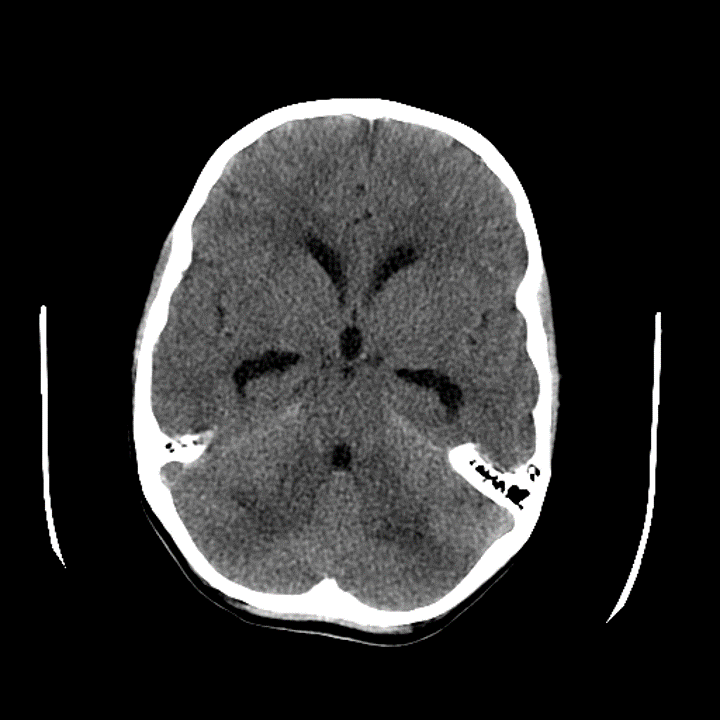
- CT
- Patchy hypoattenuation in the bilateral cerebellar hemispheres with mass effect in the posterior fossa resulting in crowding of the fourth ventricle and inferior descent of the cerebellar tonsils into the foramen magnum
- Enlargement of the lateral and third ventricles with mild rounding of the temporal horns and mild subependymal edema
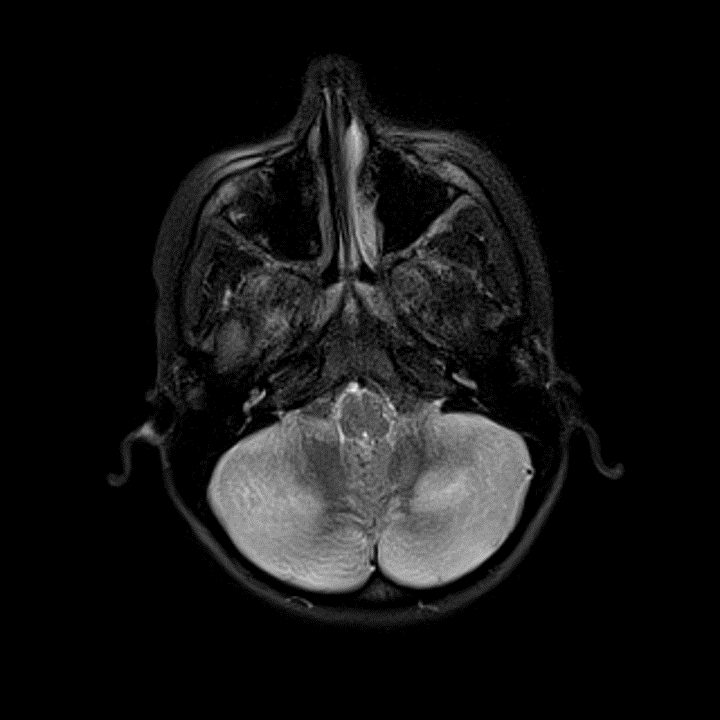
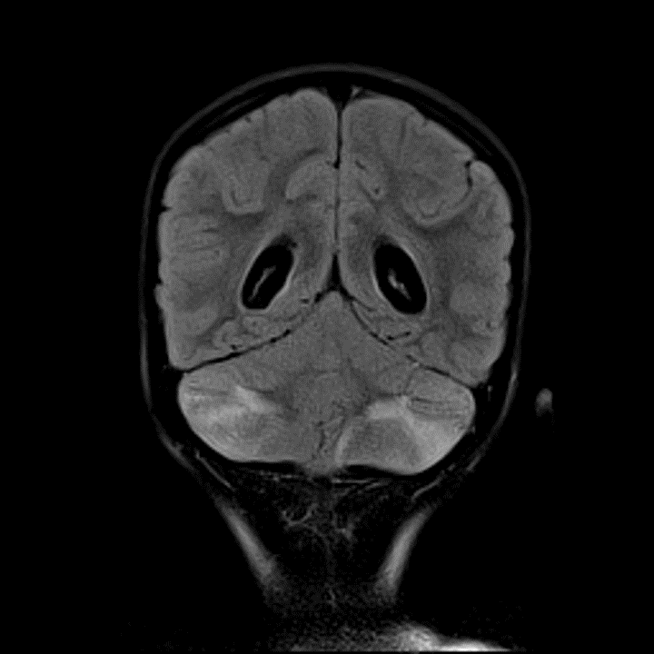
- MRI
- Patchy T2/FLAIR hyperintensity involving gray and white matter in both cerebellar hemispheres with associated cerebellar swelling and partial effacement of the fourth ventricle
- Descent of the bilateral cerebellar tonsils into the foramen magnum with mass effect on the upper cervical spinal cord
- Enlargement of the lateral and third ventricles with periventricular T2/FLAIR hyperintensity
- Thin diffuse pial enhancement along the surface of the cerebellum
- No evidence of acute infarct or hemorrhage
Diagnosis
- Cerebellitis
 Sample Report
Findings consistent with acute cerebellitis with associated cerebellar swelling resulting in mass effect on the fourth ventricle and bilateral cerebellar tonsillar herniation. Obstructive hydrocephalus with enlargement of the lateral and third ventricles with transependymal edema. Recommend urgent neurosurgical evaluation.
Thin diffuse pial enhancement along the surface of the cerebellum may be directly related to cerebellitis or a secondary effect of cerebellar edema and vascular congestion.
No evidence of acute infarct or hemorrhage.
Discussion
- Acute cerebellitis is most often postinfectious, occurring 1-2 weeks after a viral infection (e.g. varicella zoster, EBV, measles, HSV, coxsackievirus)
- It most commonly occurs in children and adolescents
- Neuroimaging is reportedly abnormal in only about 10% of cases where imaging is obtained, so the prevalence of this diagnosis on imaging largely underestimates its clinical prevalence
- Whenever there is evidence of cerebellar edema on imaging, patients need to be watched closely for development of obstructive hydrocephalus, which can be rapidly fatal
 Images
📣 Feedback?
⌨️ Keyboard Shortcuts ("K")
Help | Terms | Privacy Policy | Cookie Policy
Medical Disclaimer | © 2024 CaseStacks LLC
Related Cases
Temporarily disabled.

 View shortcuts
 Zoom/Pan
 Full screen
 Window/Level
 Expand/collapse
 Scroll
 Save the case
 Close case/tab


