Become a Pro Member for full access to this course and more.

View Plans   Log In
Expand ("E")   Collapse

Age: 11

Sex: Female

Indication: Fever, drowsiness

Save ("V")

Case #27


Findings

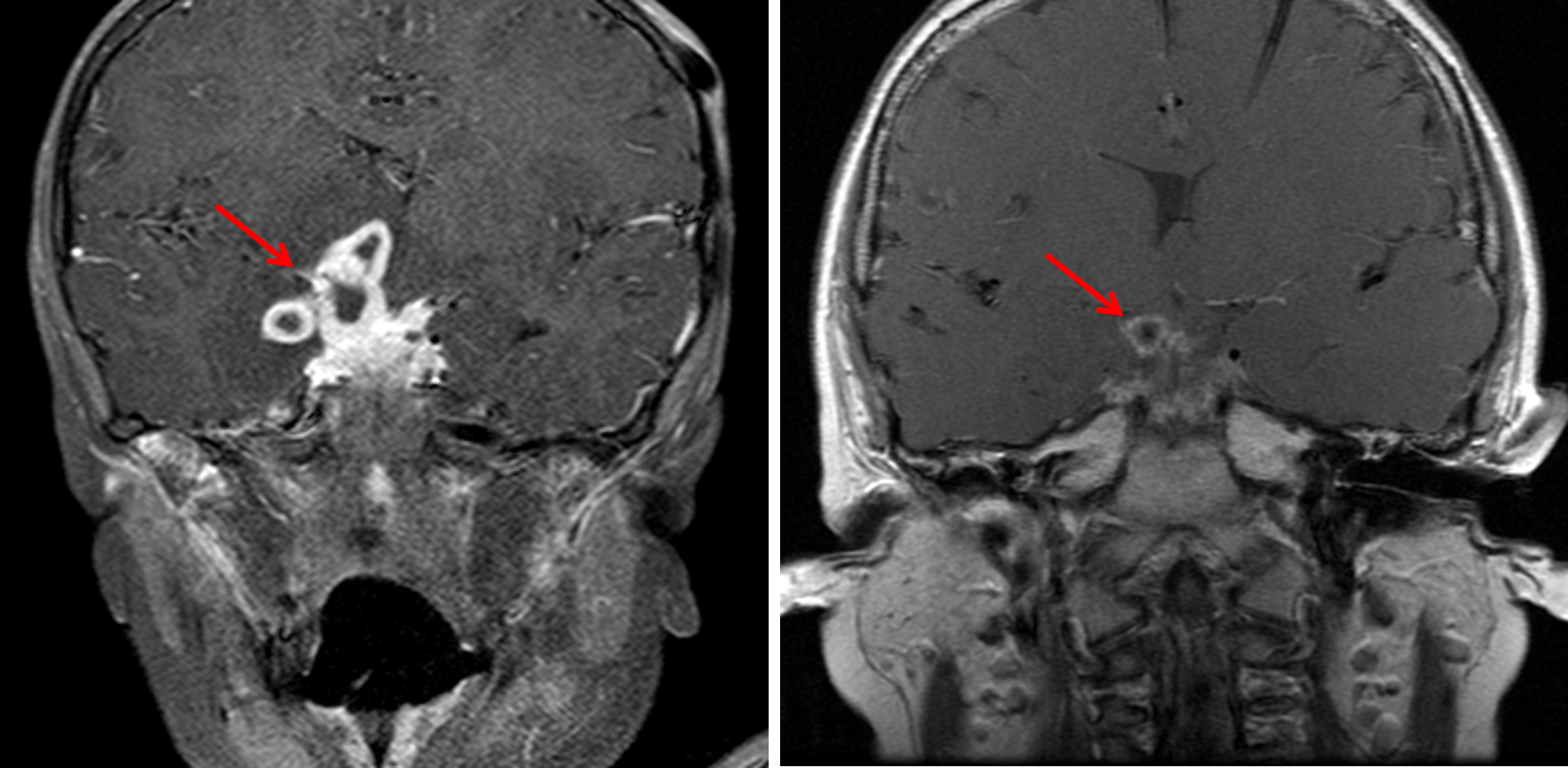
  • Nodular enhancing soft tissue filling the suprasellar cistern and extending into the cavernous sinus, interpeduncular fossa, anterior interhemispheric fissure, and right greater than left ambient cisterns and sylvian fissures with areas of internal T2 signal hypointensity
  • Peripherally enhancing components contiguously extend into the parenchyma of the inferior right frontal lobe, hypothalamus, and medial right temporal lobe with surrounding T2/FLAIR signal hyperintensity throughout a larger area of the right greater than left inferior frontal lobes, right temporal lobe, right greater than left basal ganglia and thalami, and right greater than left midbrain
  • Small area of restricted diffusion in the approximate location of the anterior commissure on the right
  • Mild generalized cerebral edema with FLAIR signal hyperintensity in multiple cerebral sulci and otherwise mild multifocal leptomeningeal enhancement
  • Abnormal enhancement about the communicating segments of the right greater than left internal carotid arteries, M1 segment of the right MCA, right posterior communicating artery, P1 segment of the right PCA, and distal basilar artery with asymmetric narrowing of the communicating segment of the right ICA
  • Left fetal-type PCA
  • Abnormal hypoenhancement of the left transverse sinus, sigmoid sinus, jugular bulb, and proximal internal jugular vein, likely related to flow-related signal loss given the appearance on other sequences
  • Left frontal approach ventricular drain with tip near the left Foramen of Monro
  • Linear area of gliosis in the right frontal lobe related to a previously removed ventricular drain
  • No acute hemorrhage, herniation, or hydrocephalus
  • Bilateral mastoid and middle ear effusions


Diagnosis

  • Tuberculous meningitis

Sample Report

Extensive basal meningitis with differential considerations including tuberculosis as well as other bacterial and fungal pathogens. Recommend CSF sampling for further evaluation.

Peripherally enhancing components contiguously extend into the parenchyma of the inferior right frontal lobe, hypothalamus, and medial right temporal lobe concerning for small abscesses with surrounding larger areas of cerebritis.

Findings concerning for vasculitis involving the right greater than left internal carotid arteries, M1 segment of the right MCA, right posterior communicating artery, P1 segment of the right PCA, and distal basilar artery with asymmetric narrowing of the communicating segment of the right ICA. A small area of restricted diffusion in the approximate location of the anterior commissure on the right could represent a small perforator infarct vs infectious debris. No additional evidence for an acute infarct.

Left frontal approach ventricular drain with tip near the left Foramen of Monro. No evidence of hydrocephalus.

Bilateral mastoid and middle ear effusions, which can be seen with otomastoiditis.


Discussion

Become a Pro Member to unlock the discussion.

View Plans   Log In


Images

Followup imaging 2.5 years after the initial presentation (on right) shows decreased bulk of enhancing soft tissue with a residual peripherally enhancing tuberculoma (red arrows).


📣 Feedback?

⌨️  Keyboard Shortcuts ("K")

Call Preparation

Prepare for call efficiently with interactive cases, sample reports, and annotated images.

1,400+ cases  91.25 CME

Call Simulator

   Neuro CT

250 cases

  Neuro MRI

146 cases

   Peds Radiographs

372 cases

   MSK Radiographs

281 cases

   Chest CT

84 cases

   Chest Radiographs

97 cases

   KUB

43 cases

   Body CT

140 cases

   Ultrasound

75 cases

Call Simulator

• Shuffle cases from our courses to simulate the mix of a call shift.
• Submit your own report before reviewing the case write-up.

Neuro Fellowship

Reviews of neuro topics with clinical pearls, differentials, and in-depth discussions.

131 cases  13.1 CME

   Brain Tumors

105 cases

   Neurodegenerative

13 cases

   Congenital Hearing Loss

13 cases

Call Preparation

   Neuro CT

250 cases

   Neuro MRI

146 cases

   Peds Radiographs

372 cases

   MSK Radiographs

281 cases

   Ultrasound

75 cases

   Chest CT

84 cases

   Chest Radiographs

97 cases

   Body CT

127 cases

   KUB

43 cases

Neuro Fellowship

   Brain Tumors

105 cases

   Neurodegenerative

13 cases

Call Simulator

• Shuffle cases from our courses.
• Submit your own report.

CME

• Earn up to 91 CME.
• Claim your CME to receive a certificate.

Call Simulator

• Shuffle cases from our courses to simulate the mix of a call shift.
• Submit your own report before reviewing the case write-up.

CME

• Earn up to 91 CME by completing cases in our radiology courses.
• Claim your CME to receive a certificate.

On Call

Quick references created specifically to help while on call.

  Peds Normals by Age

Reference database of normal imaging from birth to age 16

   Incidental Findings

Summary of consensus guidelines for managing incidental CT findings

   Media Index

Index of select illustrations & videos from our courses

  Neuro CT Mimics

Visual reference for common mimics of pathology on CT

On Call

  Peds Normals by Age

Reference database of normal imaging from birth to age 16

   Incidental Findings

Summary of consensus guidelines for managing incidental CT findings

   Media Index

Index of select illustrations & videos from our courses

  Neuro CT Mimics

Visual reference for common mimics of pathology on CT

Anatomy

Labelled radiographs and CT/MRI series teaching anatomy with a level of detail appropriate for medical students and junior residents.

   Pelvis

Pelvic MRI anatomy

   Chest

Chest radiograph & CT anatomy

   Body

Abdominal CT anatomy

   Cardiac

Cardiac CT anatomy

   Brain

Brain & calvarial anatomy on CT/MRI

   Cranial Nerves

Cranial nerves on MRI

   Shoulder

Shoulder MRI anatomy

   Knee

Knee MRI anatomy

   Temporal Bone

Resident/fellow-level anatomy

Anatomy

   Pelvis

Pelvic MRI anatomy

   Chest

Chest radiograph & CT anatomy

   Body

Abdominal CT anatomy

   Cardiac

Cardiac CT anatomy

   Brain

Brain & calvarial anatomy on CT/MRI

   Cranial Nerves

Cranial nerves on MRI

   Shoulder

Shoulder MRI anatomy

   Knee

Knee MRI anatomy

   Temporal Bone

Resident/fellow-level anatomy

Neuro Courses

Includes our call preparation neuro courses (Neuro CT and Neuro MRI) and our neuro fellowship courses.

   Neuro CT

250 cases

  Neuro MRI

146 cases

   Brain Tumors

105 cases

   Neurodegenerative

13 cases

   Congenital Hearing Loss

Coming soon

Neuro Courses

   Neuro CT

250 cases

   Neuro MRI

146 cases

   Brain Tumors

105 cases

   Neurodegenerative

13 cases

  Congenital Hearing Loss

Coming soon

CME

• Claim your CME to receive a certificate.

Call Simulator

• Shuffle cases from our courses to simulate the mix of a call shift.
• Submit your own report before reviewing the case write-up.

CME

• Earn up to 91 CME by completing cases in our radiology courses.
• Claim your CME to receive a certificate.

   Neuro CT

250 cases

   Neuro MRI

146 cases

   Peds Radiographs

372 cases

   MSK Radiographs

281 cases

   Ultrasound

75 cases

   Chest CT

84 cases

   Chest Radiographs

97 cases

   Body CT

140 cases

   KUB

43 cases

Call Simulator

• Shuffle cases from our courses to simulate the mix of a call shift.
• Submit your own report before reviewing the case write-up.

CME

• Earn up to 91 CME by completing cases in our radiology courses.
• Claim your CME to receive a certificate.

⌨️ Keyboard Shortcuts

View shortcuts

Zoom/Pan

  Full screen

  Window/Level

  Expand/collapse

  Scroll

  Save the case

  Close case/tab

Previous Slice (PC)

Next Slice (PC)

SHIFT Previous Slice (MAC)

 SHIFT Next Slice (MAC)

 Toggle presets

 Previous series (if multiple)

 Next series (if multiple)

  Previous case (⌘ ◄ on a mac)

  Next case (⌘ ► on a mac)

Toggle dark mode