Expand ("E")   Collapse

Age: 24

Sex: Female

Indication: Trauma

Save ("V")

Case #1


Findings

Chest Radiograph

  • Acute nondisplaced right first rib fracture.
  • No pleural effusion or pneumothorax.
  • Mild hazy bibasilar opacities.


CT

  • Chest
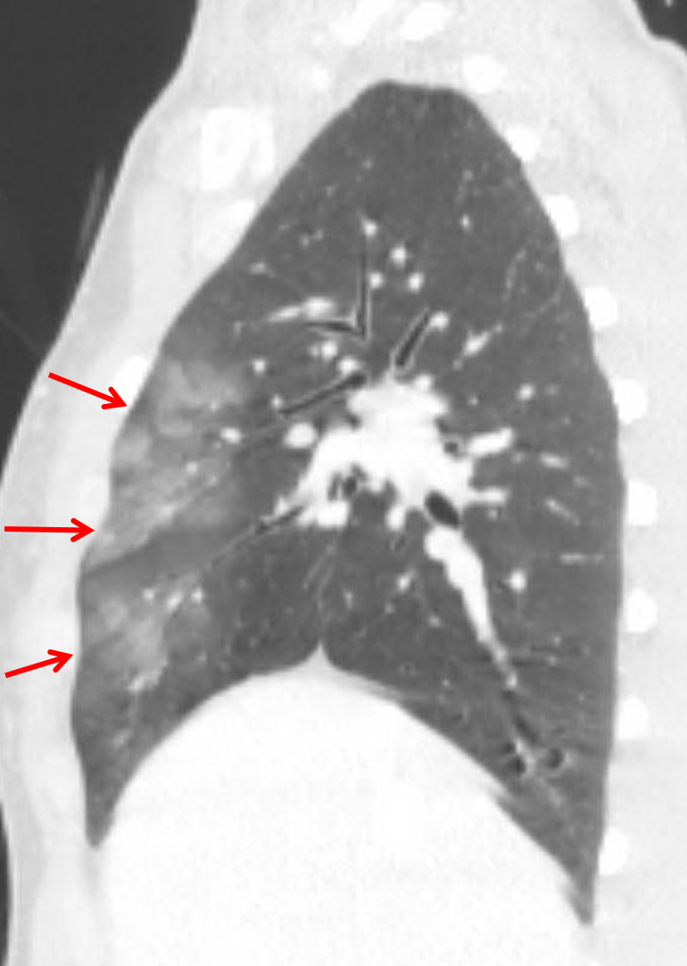
    • Mild peripheral groundglass opacities in the anterior aspects of the right upper and middle lobes and to a lesser extent in the lingula
    • Trace bilateral pneumothoraces
    • Trace anterior pneumomediastinum
    • Residual thymus in the anterior mediastinum
  • Abdomen/Pelvis
    • No acute findings
    • Narrowing and inferior deviation at the origin of the celiac artery with poststenotic dilation
  • MSK
    • Acute nondisplaced fractures of the right first rib and of the left fifth through eighth ribs
    • Acute L1 compression fracture with approximately 10% anterior height loss and no bony retropulsion
    • Possible additional acute compression fracture of the T11 vertebral body without significant height loss or bony retropulsion
    • Mild offset of the coccygeal segments


Diagnosis

  • Pulmonary contusion

Sample Report

Acute nondisplaced fractures of the right first and left fifth through eighth ribs. Trace bilateral pneumothoraces.

Trace anterior pneumomediastinum without mediastinal hematoma or evidence of aortic trauma.

Pulmonary contusion in the anterior aspects of the right upper and middle lobes and to a lesser extent in the lingula.

Acute L1 and possibly T11 compression fractures with minimal height loss and no bony retropulsion.

Age-indeterminate mild offset of the coccygeal segments which may be traumatic or developmental.

No acute traumatic findings in the abdominopelvic cavity.

Narrowing at the origin of the celiac artery likely relates to median arcuate ligament compression. Though often asymptomatic, this finding can be a cause for chronic abdominal pain.


Discussion

  • Pulmonary contusion is often located in a non-dependent area of the lung and correlates with the site(s) of blunt chest trauma
  • Pulmonary contusion will typically resolve within a few days with supportive management, so if you see groundglass opacities persisting a week or more after trauma or appearing more than 24 hours after trauma, there is likely something else going on
  • If you see bilateral dependent groundglass opacities, this is more likely to represent subsegmental atelectasis or aspiration
  • Make sure to look for associated pulmonary laceration or pneumatocele
  • In this case, the trace pneumomediastinum is likely a product of the Macklin effect: gas released from alveolar rupture tracks along bronchovascular bundles back to the mediastinum
    • However, whenever you see pneumomediastinum, make sure to look closely for evidence of tracheobronchial or esophageal injury and look for other signs of mediastinal trauma including mediastinal hematomas and aortic injuries


Images

Pulmonary contusion: groundglass opacities in the anterior aspects of the right upper and middle lobes (red arrows).


📣 Feedback?

⌨️  Keyboard Shortcuts ("K")

Call Preparation

Prepare for call efficiently with interactive cases, sample reports, and annotated images.

1,400+ cases  91.25 CME

Call Simulator

   Neuro CT

250 cases

  Neuro MRI

146 cases

   Peds Radiographs

372 cases

   MSK Radiographs

281 cases

   Chest CT

84 cases

   Chest Radiographs

97 cases

   KUB

43 cases

   Body CT

140 cases

   Ultrasound

75 cases

Call Simulator

• Shuffle cases from our courses to simulate the mix of a call shift.
• Submit your own report before reviewing the case write-up.

Neuro Fellowship

Reviews of neuro topics with clinical pearls, differentials, and in-depth discussions.

131 cases  13.1 CME

   Brain Tumors

105 cases

   Neurodegenerative

13 cases

   Congenital Hearing Loss

13 cases

Call Preparation

   Neuro CT

250 cases

   Neuro MRI

146 cases

   Peds Radiographs

372 cases

   MSK Radiographs

281 cases

   Ultrasound

75 cases

   Chest CT

84 cases

   Chest Radiographs

97 cases

   Body CT

127 cases

   KUB

43 cases

Neuro Fellowship

   Brain Tumors

105 cases

   Neurodegenerative

13 cases

Call Simulator

• Shuffle cases from our courses.
• Submit your own report.

CME

• Earn up to 91 CME.
• Claim your CME to receive a certificate.

Call Simulator

• Shuffle cases from our courses to simulate the mix of a call shift.
• Submit your own report before reviewing the case write-up.

CME

• Earn up to 91 CME by completing cases in our radiology courses.
• Claim your CME to receive a certificate.

On Call

Quick references created specifically to help while on call.

  Peds Normals by Age

Reference database of normal imaging from birth to age 16

   Incidental Findings

Summary of consensus guidelines for managing incidental CT findings

   Media Index

Index of select illustrations & videos from our courses

  Neuro CT Mimics

Visual reference for common mimics of pathology on CT

On Call

  Peds Normals by Age

Reference database of normal imaging from birth to age 16

   Incidental Findings

Summary of consensus guidelines for managing incidental CT findings

   Media Index

Index of select illustrations & videos from our courses

  Neuro CT Mimics

Visual reference for common mimics of pathology on CT

Anatomy

Labelled radiographs and CT/MRI series teaching anatomy with a level of detail appropriate for medical students and junior residents.

   Pelvis

Pelvic MRI anatomy

   Chest

Chest radiograph & CT anatomy

   Body

Abdominal CT anatomy

   Cardiac

Cardiac CT anatomy

   Brain

Brain & calvarial anatomy on CT/MRI

   Cranial Nerves

Cranial nerves on MRI

   Shoulder

Shoulder MRI anatomy

   Knee

Knee MRI anatomy

   Temporal Bone

Resident/fellow-level anatomy

Anatomy

   Pelvis

Pelvic MRI anatomy

   Chest

Chest radiograph & CT anatomy

   Body

Abdominal CT anatomy

   Cardiac

Cardiac CT anatomy

   Brain

Brain & calvarial anatomy on CT/MRI

   Cranial Nerves

Cranial nerves on MRI

   Shoulder

Shoulder MRI anatomy

   Knee

Knee MRI anatomy

   Temporal Bone

Resident/fellow-level anatomy

Neuro Courses

Includes our call preparation neuro courses (Neuro CT and Neuro MRI) and our neuro fellowship courses.

   Neuro CT

250 cases

  Neuro MRI

146 cases

   Brain Tumors

105 cases

   Neurodegenerative

13 cases

   Congenital Hearing Loss

Coming soon

Neuro Courses

   Neuro CT

250 cases

   Neuro MRI

146 cases

   Brain Tumors

105 cases

   Neurodegenerative

13 cases

  Congenital Hearing Loss

Coming soon

CME

• Claim your CME to receive a certificate.

Call Simulator

• Shuffle cases from our courses to simulate the mix of a call shift.
• Submit your own report before reviewing the case write-up.

CME

• Earn up to 91 CME by completing cases in our radiology courses.
• Claim your CME to receive a certificate.

   Neuro CT

250 cases

   Neuro MRI

146 cases

   Peds Radiographs

372 cases

   MSK Radiographs

281 cases

   Ultrasound

75 cases

   Chest CT

84 cases

   Chest Radiographs

97 cases

   Body CT

140 cases

   KUB

43 cases

Call Simulator

• Shuffle cases from our courses to simulate the mix of a call shift.
• Submit your own report before reviewing the case write-up.

CME

• Earn up to 91 CME by completing cases in our radiology courses.
• Claim your CME to receive a certificate.

⌨️ Keyboard Shortcuts

View shortcuts

Zoom/Pan

  Full screen

  Window/Level

  Expand/collapse

  Scroll

  Save the case

  Close case/tab

Previous Slice (PC)

Next Slice (PC)

SHIFT Previous Slice (MAC)

 SHIFT Next Slice (MAC)

 Toggle presets

 Previous series (if multiple)

 Next series (if multiple)

  Previous case (⌘ ◄ on a mac)

  Next case (⌘ ► on a mac)

Toggle dark mode