Expand ("E")   Collapse

Age: 18

Sex: Female

Indication: Trauma, no fracture on cervical spine CT

Save ("V")

Case #1


Findings

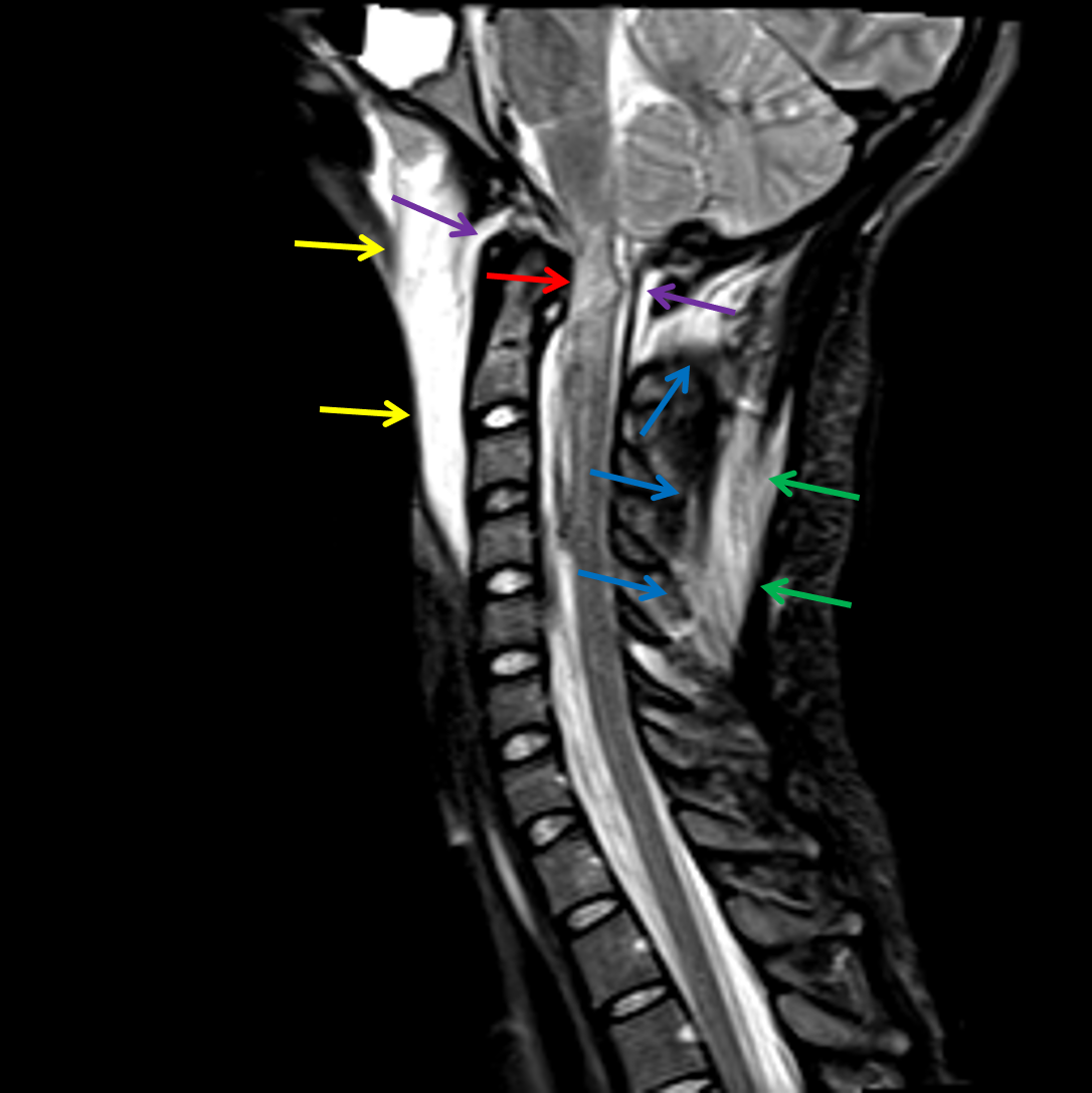
  • T2/STIR signal hyperintensity in the anterior and posterior atlantooccipital membrane and bilateral atlantooccipital joints with mild widening of both atlantooccipital joints
  • Widening of the basion-dens interval with discontinuity of the apical ligament and mixed signal material about the odontoid process and in the supraodontoid space exerting mass effect on the upper cervical spinal cord at the level of C1
  • No discontinuity of the tectorial membrane
  • Abnormal T2/STIR signal hyperintensity extending from the caudal medulla through the cervical spinal cord to the level of C2-C3, most pronounced at the level of C1
  • T2/STIR signal hyperintensity in the interspinous ligament at multiple levels, most pronounced at C5-C6
  • T2/STIR signal hyperintensity in the nuchal ligament extending from the occiput inferiorly to the level of C6
  • T2/STIR signal hyperintensity in the prevertebral soft tissues extending from the skull base through the level of C4-C5
  • Large T1 isointense, T2 hyperintense epidural collection extending from the level of C2 inferiorly through the cervical and visualized upper thoracic spine. This collection is mainly located ventrally in the cervical spine and dorsally in the upper thoracic spine
  • Partially imaged endotracheal and enteric tubes
  • Right greater than left pleural effusions


Diagnosis

  • Atlantooccipital dissociation

Sample Report

Findings consistent with atlantooccipital dissociation with disruption of the apical ligament and concern for bilateral C1-C2 joint capsular injury. Mild associated widening of the basion-dens interval and bilateral C1-C2 articulations. The tectorial membrane appears intact.

Extensive ligamentous injury involving the anterior and posterior atlantooccipital membranes as well as the interspinous and nuchal ligaments at multiple levels.

Hemorrhage about the odontoid process with cord contusion at the level of C1. Surrounding edema extends from the caudal medulla to the level of C2-C3.

Large epidural hematoma extending from C2 through the upper thoracic spine, incompletely evaluated on this study, which results in mild multilevel narrowing of the spinal canal. Consider dedicated thoracic and lumbar spine MRI for a more complete assessment of this collection.

Extensive prevertebral edema/hematoma.

No findings to suggest an acute fracture.


Discussion

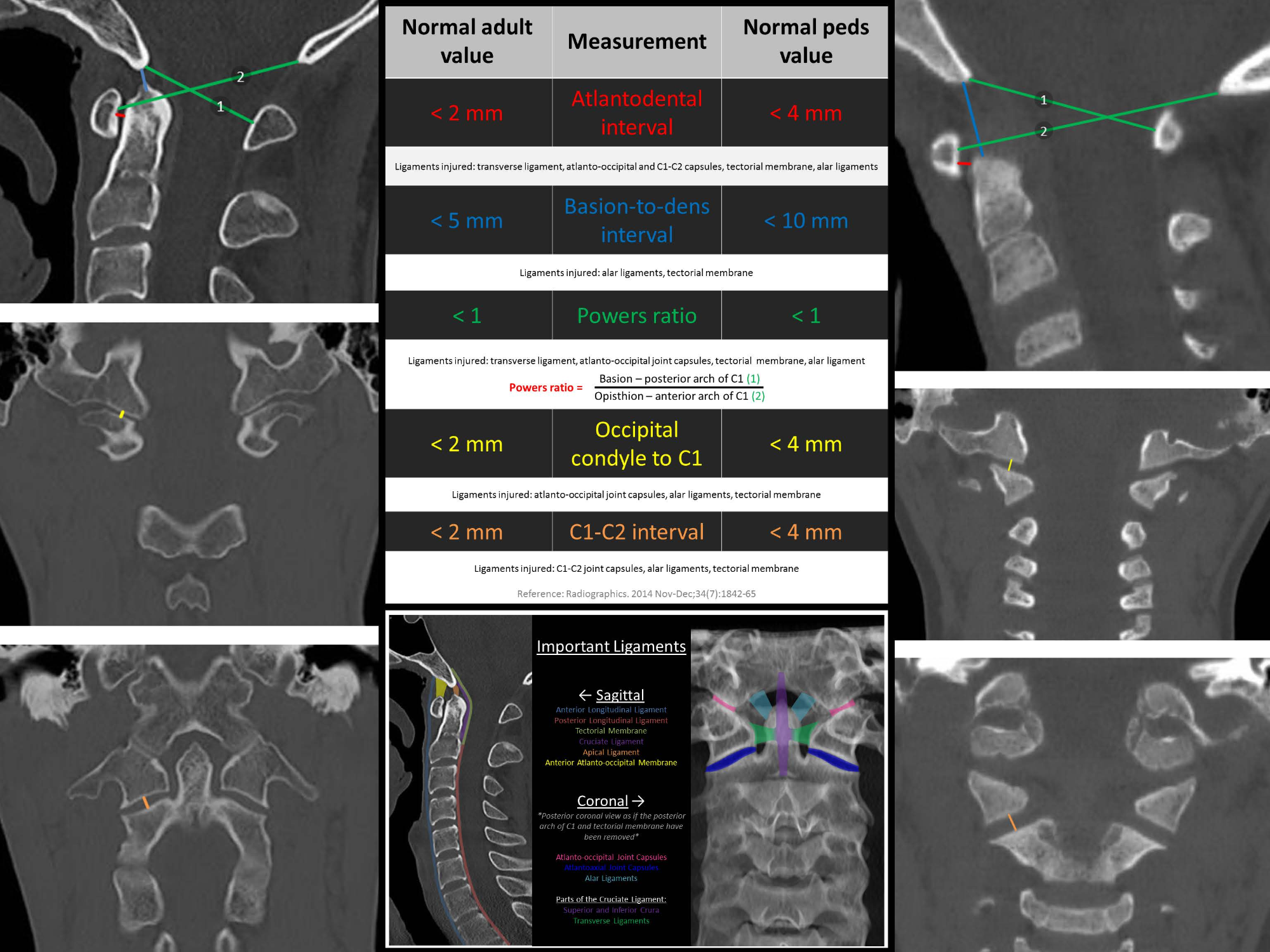
  • The atlantooccipital joint is commonly injured in high impact trauma, particularly in children, and though serious often has only subtle associated findings on CT
  • Make sure to assess the intervals shown in the annotated images below in trauma cases to avoid missing these injuries
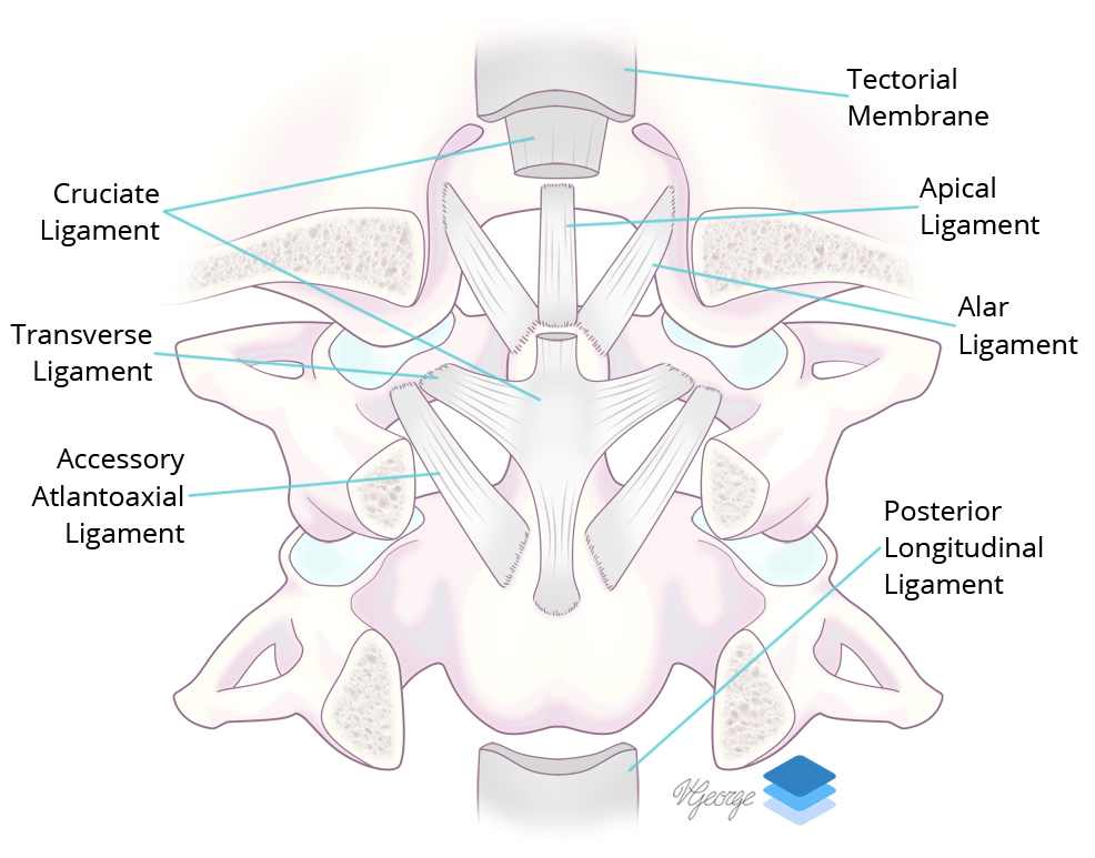
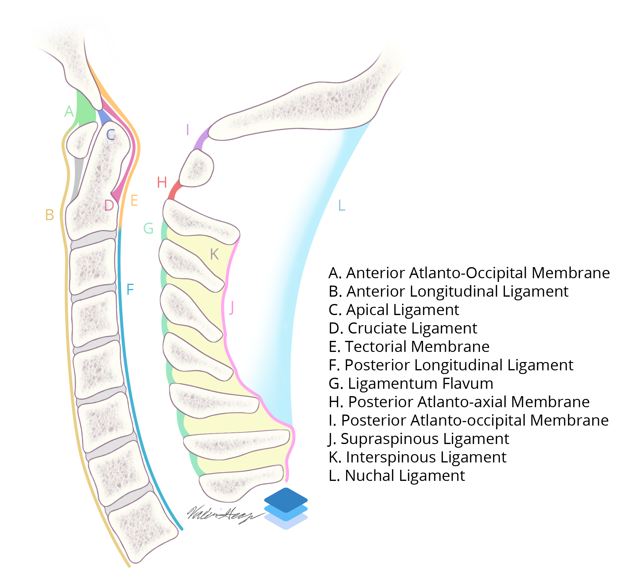
  • Many of the stabilizing ligaments can be identified and assessed on MRI, which are shown in the annotated images below
  • The two most important ligaments for stabilizing the craniocervical junction are the alar ligaments (which extend obliquely from the dens to the occipital condyles) and the tectorial membrane (the superior continuation of the posterior longitudinal ligament)


Images

Atlantooccipital dissociation: Red arrow: upper cervical cord and medullary contusion. Purple arrows: edema in the anterior and posterior atlantooccipital membranes. Yellow arrows: prevertebral edema/hematoma. Blue arrows: edema in the interspinous ligament. Green arrows: edema in the nuchal ligament. Coronal view of the major ligaments at the craniocervical junction. Sagittal illustration of the major cervical spine ligaments. Diagram of helpful measurements to know.


📣 Feedback?

⌨️  Keyboard Shortcuts ("K")

Call Preparation

Prepare for call efficiently with interactive cases, sample reports, and annotated images.

1,400+ cases  91.25 CME

Call Simulator

   Neuro CT

250 cases

  Neuro MRI

146 cases

   Peds Radiographs

372 cases

   MSK Radiographs

281 cases

   Chest CT

84 cases

   Chest Radiographs

97 cases

   KUB

43 cases

   Body CT

140 cases

   Ultrasound

75 cases

Call Simulator

• Shuffle cases from our courses to simulate the mix of a call shift.
• Submit your own report before reviewing the case write-up.

Neuro Fellowship

Reviews of neuro topics with clinical pearls, differentials, and in-depth discussions.

131 cases  13.1 CME

   Brain Tumors

105 cases

   Neurodegenerative

13 cases

   Congenital Hearing Loss

13 cases

Call Preparation

   Neuro CT

250 cases

   Neuro MRI

146 cases

   Peds Radiographs

372 cases

   MSK Radiographs

281 cases

   Ultrasound

75 cases

   Chest CT

84 cases

   Chest Radiographs

97 cases

   Body CT

127 cases

   KUB

43 cases

Neuro Fellowship

   Brain Tumors

105 cases

   Neurodegenerative

13 cases

Call Simulator

• Shuffle cases from our courses.
• Submit your own report.

CME

• Earn up to 91 CME.
• Claim your CME to receive a certificate.

Call Simulator

• Shuffle cases from our courses to simulate the mix of a call shift.
• Submit your own report before reviewing the case write-up.

CME

• Earn up to 91 CME by completing cases in our radiology courses.
• Claim your CME to receive a certificate.

On Call

Quick references created specifically to help while on call.

  Peds Normals by Age

Reference database of normal imaging from birth to age 16

   Incidental Findings

Summary of consensus guidelines for managing incidental CT findings

   Media Index

Index of select illustrations & videos from our courses

  Neuro CT Mimics

Visual reference for common mimics of pathology on CT

On Call

  Peds Normals by Age

Reference database of normal imaging from birth to age 16

   Incidental Findings

Summary of consensus guidelines for managing incidental CT findings

   Media Index

Index of select illustrations & videos from our courses

  Neuro CT Mimics

Visual reference for common mimics of pathology on CT

Anatomy

Labelled radiographs and CT/MRI series teaching anatomy with a level of detail appropriate for medical students and junior residents.

   Pelvis

Pelvic MRI anatomy

   Chest

Chest radiograph & CT anatomy

   Body

Abdominal CT anatomy

   Cardiac

Cardiac CT anatomy

   Brain

Brain & calvarial anatomy on CT/MRI

   Cranial Nerves

Cranial nerves on MRI

   Shoulder

Shoulder MRI anatomy

   Knee

Knee MRI anatomy

   Temporal Bone

Resident/fellow-level anatomy

Anatomy

   Pelvis

Pelvic MRI anatomy

   Chest

Chest radiograph & CT anatomy

   Body

Abdominal CT anatomy

   Cardiac

Cardiac CT anatomy

   Brain

Brain & calvarial anatomy on CT/MRI

   Cranial Nerves

Cranial nerves on MRI

   Shoulder

Shoulder MRI anatomy

   Knee

Knee MRI anatomy

   Temporal Bone

Resident/fellow-level anatomy

Neuro Courses

Includes our call preparation neuro courses (Neuro CT and Neuro MRI) and our neuro fellowship courses.

   Neuro CT

250 cases

  Neuro MRI

146 cases

   Brain Tumors

105 cases

   Neurodegenerative

13 cases

   Congenital Hearing Loss

Coming soon

Neuro Courses

   Neuro CT

250 cases

   Neuro MRI

146 cases

   Brain Tumors

105 cases

   Neurodegenerative

13 cases

  Congenital Hearing Loss

Coming soon

CME

• Claim your CME to receive a certificate.

Call Simulator

• Shuffle cases from our courses to simulate the mix of a call shift.
• Submit your own report before reviewing the case write-up.

CME

• Earn up to 91 CME by completing cases in our radiology courses.
• Claim your CME to receive a certificate.

   Neuro CT

250 cases

   Neuro MRI

146 cases

   Peds Radiographs

372 cases

   MSK Radiographs

281 cases

   Ultrasound

75 cases

   Chest CT

84 cases

   Chest Radiographs

97 cases

   Body CT

140 cases

   KUB

43 cases

Call Simulator

• Shuffle cases from our courses to simulate the mix of a call shift.
• Submit your own report before reviewing the case write-up.

CME

• Earn up to 91 CME by completing cases in our radiology courses.
• Claim your CME to receive a certificate.

⌨️ Keyboard Shortcuts

View shortcuts

Zoom/Pan

  Full screen

  Window/Level

  Expand/collapse

  Scroll

  Save the case

  Close case/tab

Previous Slice (PC)

Next Slice (PC)

SHIFT Previous Slice (MAC)

 SHIFT Next Slice (MAC)

 Toggle presets

 Previous series (if multiple)

 Next series (if multiple)

  Previous case (⌘ ◄ on a mac)

  Next case (⌘ ► on a mac)

Toggle dark mode